Anti-Elevation Syndrome
Last updated: October 2026
Also known as "J" deformity of IO.
Placement of the posterior fibres at, or anterior to, the inferior rectus insertion produces a “J” deformity, and will limit elevation of the eye. The complication of limited elevation has been termed by Mims et al as anti-elevation syndrome.
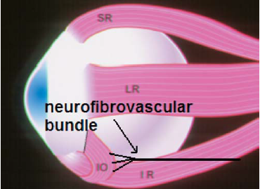
Stager has shown, through anatomical dissections, that the neurofibrovascular bundle of the inferior oblique inserts into the posterior fibres and can act as a tether to hold the eye down. He found that the ligamentous structure of the neurofibrovascular bundle provides the ancillary origin for the posterior temporal fibres of the inferior oblique muscle when its insertion is transposed anteriorly. Because of these concerns, the full anteriorization with a “J” deformity should be reserved for severe bilateral DVD with inferior oblique overaction. Mims stated that the author’s procedure of keeping the posterior fibres posterior to the inferior rectus insertion prevents this complication.
Drawing shows the “J” deformity anteriorization and the neurofibrovascular bundle inserting into the posterior fibres of the inferior oblique muscle. When the posterior muscle fibres are moved anterior to the inferior rectus insertion the neurofibrovascular bundle tethers the eye, resulting in limited elevation.
Inferior oblique weakening procedures compared
| Procedure | What is done | Best for | Watch out for |
|---|---|---|---|
| Recession | Insertion moved back (e.g. 8–14 mm) towards the IR | Mild to moderate IO overaction | Under-correction; adherence syndrome if fat is breached |
| Myectomy / disinsertion | A segment of the muscle is excised or the insertion cut | Moderate overaction; quick and reliable | Reattachment, bleeding from the vortex vein |
| Anterior transposition | Insertion placed beside the lateral end of the IR insertion | IO overaction with DVD | Anti-elevation syndrome if posterior fibres go forward of the IR (J deformity) |
| Denervation and extirpation | Nerve to the IO cut and muscle removed | Severe or recurrent overaction | Irreversible; pupil changes (ciliary ganglion nearby) |
Grade IO overaction on side gaze before choosing (see the oblique overaction grading). Inspect the inferotemporal quadrant for vortex veins during any IO procedure.
Revise with flip cards
Key sources
- Mims JL 3rd, Wood RC. Bilateral anterior transposition of the inferior obliques. Arch Ophthalmol. 1989;107:41-44
- Stager DR. The neurofibrovascular bundle of the inferior oblique muscle as its ancillary origin. Trans Am Ophthalmol Soc. 1996;94:1073-1094
📘 Want the exam-ready deep dive? This topic, and much more is covered in Ophthalmology Explorer (A High-Yield Clinical Reference for Ophthalmology Residents, Fellows & Board Exams) — available on Kindle. See all my books.
How to cite this page
Patel D. Anti-Elevation Syndrome. RPC Rounds (Sadbhaav Eye Clinic). Updated October 2026. Available at: https://www.sadbhaavclinic.com/aes.html. Accessed [date].
Want to know more about treatment? Read about Squint Treatment at Sadbhaav or book a consultation with Dr. Dhaval Patel.