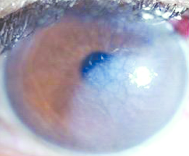
Descemet’s membrane detachment classification
Last updated: September 2026
Descemet membrane detachment (DMD) is a potentially vision-threatening complication seen most often after cataract surgery, though it can follow any intraocular procedure. Prompt recognition matters — untreated DMD can progress to permanent corneal decompensation requiring keratoplasty, while most cases resolve fully with timely, appropriately-targeted management.
Anatomy: it's not always just the DM
A useful modern refinement: what detaches is not always the Descemet membrane alone. The pre-Descemet layer (PDL) — also called Dua's layer — lies just anterior to the DM and can detach together with it or separately. This distinction affects both appearance on imaging and prognosis:
- Type 1: PDL and DM detach together, remaining attached to each other
- Type 2: DM detaches alone, separate from the PDL
- Mixed type: both patterns are present in different areas of the same detachment
Diagnosis: AS-OCT is now central
Anterior segment optical coherence tomography (AS-OCT) is the primary tool for detecting DMD and mapping its extent — it distinguishes true DMD from simple corneal edema, characterizes the type of detachment, and guides the decision between observation and intervention. Have a high index of suspicion whenever corneal edema after surgery is unexplained or has an atypical, localized pattern rather than diffuse stromal haze.
Classification and management (Jacob-Agarwal system)
The most widely used clinico-tomographic classification, still current in the 2024 literature, groups DMD into four types based on etiopathogenesis, clinical features, and ASOCT findings:
| Type | Causal elements | Management |
|---|---|---|
| Rhegmatogenous | Hole, tear, dialysis in the DM | Small, clinically insignificant: Observation Clinically significant: Pneumodescemetopexy + supradescemetic fluid drainage + post-op positioning |
| Tractional | Incarceration in synechiae, wound, suture, or graft-host junction; long-standing detachment adhering to intraocular contents with contraction | Small, clinically insignificant: Observation Clinically significant, healthy endothelium: Relaxing descemetotomy + pneumodescemetopexy + supradescemetic fluid drainage + post-op positioning Unhealthy endothelium: Penetrating or endothelial keratoplasty |
| Bullous | Viscoelastic separation, trapped blood, infection, inflammation, Anwar's big bubble technique, trauma | Intraoperative nick or YAG descemetopuncture, tailored to cause |
| Complex | Poorly repositioned detachment — including DMEK graft post-op re-detachments, or long-standing rhegmatogenous detachment with fibrosis in combination with other patterns | Observation, refloatation, or endothelial/penetrating keratoplasty depending on endothelial viability |
General management principles
- First-line: Descemetopexy with air or gas (SF6/C3F8) is the initial treatment of choice for most clinically significant detachments
- Suture fixation can be added when the detachment is large or air/gas alone fails to appose the membrane
- Non-responsive cases — those with persistent detachment, unhealthy endothelium, or long-standing fibrotic change — may ultimately need endothelial keratoplasty (DSEK/DMEK) or, rarely, penetrating keratoplasty
- Early intervention matters: prolonged detachment risks irreversible endothelial cell loss and permanent stromal scarring
Key references: Beniwal A, Vanathi M, et al. Descemet's membrane detachment: an updated comprehensive review. Indian J Ophthalmol. 2024;72(11):1560-1568. | Jacob S, Agarwal A, et al. A new clinico-tomographic classification and management algorithm for DMD. Cont Lens Anterior Eye. 2015;38:327-333.
📘 Want the exam-ready deep dive? This topic is covered in iNotes 2027: Cornea and Refractive (Ophthalmology Postgraduate Exam Notes) — available on Kindle. See all my books.
Want to know more about treatment? Read about Corneal Topography at Sadbhaav or book a consultation with Dr. Dhaval Patel.