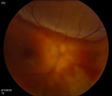
High IOP in Retinal Detachment
Last updated: September 2026
Retinal detachment classically presents with low intraocular pressure — fluid pumped through the detached retina increases uveoscleral outflow. Elevated IOP in the setting of retinal detachment is the exception, not the rule, and should immediately raise suspicion for Schwartz-Matsuo syndrome rather than being dismissed as an unrelated finding.
Schwartz-Matsuo syndrome: the classic triad
- Rhegmatogenous retinal detachment
- Elevated intraocular pressure, often with marked diurnal fluctuation
- Anterior chamber cells in an open angle, without other signs of active uveitis
Mechanism
Photoreceptor outer segments escape through the retinal break into the vitreous and anterior chamber, where they mechanically obstruct trabecular meshwork outflow. This was confirmed by electron microscopy of aqueous samples showing photoreceptor outer segments — a useful diagnostic clue when the presentation is atypical. Critically, the anterior chamber cells in this condition do not respond to topical corticosteroids, unlike true uveitis — a practical bedside clue when the diagnosis is in doubt.
Clinical clues that should trigger suspicion
- Elevated IOP in an eye with open angles and minimal-to-moderate anterior chamber reaction that doesn't fit typical anterior uveitis
- History of ocular trauma, recent intraocular surgery, floaters, photopsia, or peripheral visual field loss
- IOP that fluctuates significantly through the day
- Steroid-unresponsive anterior chamber cell
A dilated peripheral retinal exam is essential in any patient with unexplained unilateral IOP elevation and atypical anterior chamber reaction — missing an underlying retinal break/detachment delays the definitive treatment and risks permanent visual loss.
Management
- Definitive treatment is retinal detachment repair (scleral buckle, pars plana vitrectomy with endolaser and gas tamponade, or a combination) — this is curative for the syndrome, not just the detachment
- IOP is controlled in the interim with topical antihypertensives and oral carbonic anhydrase inhibitors (e.g., acetazolamide) while awaiting definitive repair
- Topical pilocarpine may help open trabecular meshwork pores, though this use is off-label
- IOP and anterior chamber cells typically normalize promptly once the retinal detachment is successfully repaired — a favorable prognostic feature that further supports the diagnosis retrospectively
Key references: Schwartz-Matsuo Syndrome, EyeWiki. | StatPearls, Schwartz-Matsuo Syndrome, 2023. | Schwartz-Matsuo syndrome: an important cause of secondary glaucoma. Am J Ophthalmol Case Rep. 2020.
📘 Want the exam-ready deep dive? This topic, and much more is covered in Ophthalmology Explorer (A High-Yield Clinical Reference for Ophthalmology Residents, Fellows & Board Exams) — available on Kindle. See all my books.
Want to know more about treatment? Read about Retina Treatment at Sadbhaav or book a consultation with Dr. Dhaval Patel.