Iris Nodules and Spots: Koeppe, Busacca, Lisch and More
Last updated: October 2026
Iris nodules and spots are a favourite spot-diagnosis. The location, colour and whether both eyes are involved usually give the answer.
Inflammatory nodules
| Nodule | Location and look | Association |
|---|---|---|
| Koeppe nodules | Small, translucent, at the pupil margin | Granulomatous uveitis (occasionally non-granulomatous) |
| Busacca nodules | On the iris surface away from the pupil; larger, gelatinous | Granulomatous uveitis: sarcoid, TB, syphilis, VKH |
| Iris roseola / papulae | Dilated vascular tufts or pink nodules | Secondary syphilis |
| Iris pearls | Small white chalky beads near the pupil | Lepromatous leprosy |
| Iris granuloma | Larger fleshy mass | Sarcoidosis, TB, fungal infection |
Developmental, pigmented and other lesions
| Lesion | Look | Association |
|---|---|---|
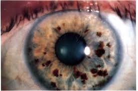
| Lisch nodules | Bilateral, smooth, tan dome-shaped melanocytic hamartomas; increase with age | Neurofibromatosis type 1 (a diagnostic criterion) |
| Juvenile xanthogranuloma | Yellow-orange vascular nodule | Infants; spontaneous hyphaema and glaucoma; skin lesions |
| Brushfield spots | Grey-white spots ringing the mid-peripheral iris | Down syndrome (also some normal light irides, where they are called Kunkmann–Wolffian bodies) |
| Iris mammillations | Tiny regular villous elevations on a dark iris | Oculodermal melanocytosis, phakomatosis pigmentovascularis |
| Iris naevus | Flat or slightly raised pigmented lesion | Common; photograph and follow up |
| Iris melanoma | Growth, prominent feeder vessels, ectropion uveae, raised IOP, seeding (ABCDEF: Age young, Blood, Clock hour inferior, Diffuse, Ectropion, Feathery margins) | Refer to ocular oncology |
| Iris cyst | Pigment epithelial (smooth, at the pupil) or stromal (translucent) | Usually benign; iris epithelial cysts can mimic plateau iris |
Where residents slip
- Calling Busacca nodules pathognomonic: they are characteristic of granulomatous uveitis but not specific to one cause.
- Missing JXG in an infant with a spontaneous hyphaema.
- Confusing Lisch nodules (bilateral hamartomas, NF1) with iris naevi.
Related: Uveitis made easy, Uveitis work-up, Ocular tumours.
Revise with flip cards
📘 Want the exam-ready deep dive? This topic is covered in iNotes 2027: Basic Science & Optics (Ophthalmology Postgraduate Exam Notes) — available on Kindle. See all my books.
Experiencing symptoms like these? Learn about our Eye Department or book a consultation with Dr. Dhaval Patel.