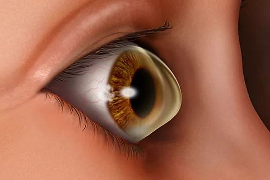
✦Keratoconus is a
noninflammatory , ectatic corneal condition characterized by central or
paracentral stromal thinning , apical protrusion and irregular astigmatism
✦British physician, Jhon
Nottingham in 1854 did practical observations on
conical cornea
✦50-230
/ 100000individuals
✦M=F
✦Starts at puberty, over a
period of 10 to 20 years the process continues until the progression gradually
stops
✦Familial
incidence= 65%, Autosomal dominant with variable penetrance
✦Pathophysiology:
✤Antioxidant
deficiency
✤Proteinase
and antiproteinase imbalance: up-regulation of
degradative enzymes and the down-regulation of proteinase inhibitors could
result in a degradation of the extracellular matrix of the stroma
✤Apoptosis: Keratocytes from keratoconus corneas have been found to have four times the interleukin-1 binding sites, when compared to nonkeratoconus corneas. This may result in an
increased sensitivity of the keratocytes in keratoconus to the effects of
interleukin-1. Interleukin-1 has also been shown to induce apoptosis or
controlled cell death of stromal keratocytes in vitro.
✤Contact
lens wear is another form of corneal microtrauma:
17.5% to 26.5%
✤ectodermal
disease, then associations with atopic disease and
tapetoretinal degenerations
✦Pathology:
✤Breaks
in the epithelial layer can be associated with
epithelium growing posteriorly into Bowman's layer and collagen growing
anteriorly into the epithelium, forming Z-shaped interruptions at the level of
Bowman's layer. These Z-shaped areas are typical of keratoconus.
✤Fleischer
ring found at the base of the cone
✤normal-sized
collagen fibers; however, the number of collagen
lamellae was abnormally low. The number found within the cone was less than
half (41%) the number outside of the cone.
✤Endothelial
cell pleomorphism and polymegathism occur in
keratoconus
✦Clinical
Features:
✤Late teens
✤Blurring of vision
✤Shadowing around images
✤Glare, halos, ocular irritation
✤Frequent changes in spectacle
number
✤Contrast sensitivity measurement
may, however, uncover visual dysfunction before Snellen visual acuity loss can
be measured
✤Two types of cones have been
described. The round or nipple-shaped cone is
smaller in diameter, while the larger oval or sagging cone may extend to the limbus and is more prone to contact lens fitting
problems.
✦Signs:
✤Irregular astigmatism
✤Striae occur in the posterior
stroma, just anterior to Descemet's membrane.
✤Red reflex Oil droplet sign
✤Scissoring reflex
✤Vogt ‘s straie
✤Fleischer’s ring
✤Prominent corneal nerves
✤Corneal topograph
✤Progressive corneal thinning
✤Munson’s sign
✤Central corneal scarring: Factors
predictive of incident corneal scarring include corneal curvature greater than
52 diopters (D), contact lens wear, corneal staining, and age less than 20
years.
✦From
Fruste Keratoconus (FFKC) was originally described
by Prof. Marc Amsler (1891-1961) based on reflection Placidodisk photography,
prior to the development of computerized corneal imaging technologies. FFKC was
used to describe an abortive form of the disease that may progress or may not.
✦Investigations:
✤The keratometer is an invaluable, widely available tool for measuring corneal
curvature. Inability to superimpose the central keratometric rings suggests
irregular corneal astigmatism, a hallmark of keratoconus.
✤Keratoscopy
or videokeratography, based on the Placido disk,
can provide qualitative contour information. In early keratoconus, a focal area
of increased corneal curvature appears as an isolated area of smaller ring
spacing and distortion. As the condition progresses, the ring spacing decreases
overall and becomes increasingly irregular
✦Rabinowitz has suggested four quantitative videokeratographic indices as an aid for screening patients for
keratoconus. These indices include
1.Central corneal
power value greater than 47.2 D
2.Inferior–superior
dioptric asymmetry (I-S value) over 1.2
3.Sim-K
astigmatism greater than 1.5 D
4.Skewed
radial axes (SRAX) greater than 21 degrees.
✦Indices
✤Simulated keratometry (SimK)
✤Surface asymmetry index (SAI):
✤Asymmetric bow tie (AB) with
skewed radial axes (SRAX): Skewing of more than 30° is described as
significantly abnormal
✤Rabinowitz/Mc
Donnel diagnostic criteria consists of two
topography derived indices, which are as follows;
▪Central K-value > 47.20 D
and
▪Inferior-Superior asymmetry
(I-S value) > 1.4 D
✤Rabinowitz/Rasheed’s described KISA% index:
▪Uses 4 parameters →
▪Keratometry; I-S value; the AST
index, which quantifies the degree of regular corneal astigmatism (simulated
flat and steep keratometry values, Sim K1 and Sim K2); and SRAX, which is an
expression of irregular astigmatism.
▪KISA% > 100% is considered
as highly suggestive of keratoconus.
✤Keratoconus-prediction
index(KPI) → Indices of Maeda and
Klyce
▪Derived from eight other quantitative
videokeratographic indices.
▪Two simulated K values (steep
and flat powers), differential sector index (DSI), center/surround index (CSI),
opposite sector index (OSI), surface asymmetry index (SAI), analyzed area (AA),
and the irregular astigmatism index (IAI).
Amsler-Krumeich Classification for Keratoconus
Classification based on mean K-readings on the anterior curvature sagittal map, thickness at the thinnest location, and the refractive error of the patient.
STAGE
FINDINGS
1
Eccentric steepening
Myopia, induced astigmatism, or both <5.00 D
Mean central K readings <48 D
2
Myopia, induced astigmatism, or both from 5.00 to 8.00 D
Mean central K readings <53.00 D Abscence of scarring Corneal thickness >400 micron
3
Myopia, induced astigmatism, or both from 8.00 to 10.00 D
Mean central K readings >53.00 D
Abscence of scarring
Corneal thickness 300 – 400 micron
4
Refraction not measurable
Mean central K readings >55.00 D
Central corneal scarring
Corneal thickness < 200 micron
Keratoconus Severity Score (KSS) Ranking Scheme
Grade
Stage
Corneal scarring*
Slit-lamp signs*
Axial Pattern
Other Features
0
Normal topography
None
None
Typical
Average corneal power (ACP) ≤ 47.75 D, Higher-order RMS error** ≤ 0.65
1
Atypical Topography
None
None
Atypical:
- Irregular
-Sup. bowie
-Inf. bowie
-Inf. or Sup. area of steepening no more than 3.00 D steeper than ACP
ACP ≤ 48.00 D, Higher-order RMS error ≤ 1.00
2
Suspect Topography
None
None
Isolated area of steepening:
-Inferior
-Superior
-Central steep
Additional features: ACP ≤ 49.00 D or Higher-order RMS error > 1.00, ≤ 1.50
3
Mild disease
None
Possible
Consistent with KCN
Additional features: ACP ≤ 52.00 D or Higher-order RMS error > 1.50, ≤ 3.50
4
Moderate disease
Add features: Corneal scarring and overall CLEK grade up to 3.0
Possible
Consistent with KCN
Additional features: ACP > 52.00 D, ≤ 56.00 D or Higher-order RMS error > 3.50, ≤ 5.75
5
Severe disease
Add features: Corneal scarring CLEK grade 3.5 or greater overall
Must have
Consistent with KCN
Additional features: ACP > 56.00 D or Higher-order RMS error > 5.75"
*consistent with KCN
**higher-order first corneal surface wavefront root mean square (RMS) error
For grades 0-1, all of the parameters in a category must be met. For all grades, the required features must be met. The worst of the additional features is then assessed, with the “worst” of the features carrying the greater weight (as long as the required features are met).
✦ABCD
Classification: The ABCD classification is measured
at the cone.
✤A: Anterior radius of curvature
from a 3.0-mm zone centered on thinnest point
✤B: Posterior (back) radius of
curvature from a 3.0-mm zone centered on the thinnest point
✤C: Minimal corneal thickness
(not apical)
✤D: Best spectacle-corrected
visual acuity
✦Systemic
Association:
✤ATOPY
▪Asthma
▪Atopickeratoconjunctivitis
▪Hay fever
▪Eczema
✤CONNECTIVE TISSUE DISORDERS
▪Marfan’s syndrome: An increased
prevalence (38%[20] to 58%) of mitral valve prolapse has been found in
keratoconus patients
▪EDS
▪Osteogenesis imperfecta
✤MISCELLANEOUS
▪Down’s: 5.5% and 15%
‣structural or biochemical
changes
‣habitual eye rubbing
▪Turner’s syndrome
✤Diabetes
offered a protective effect regarding keratoconus. (also smoking?? As they
cause C3R like effect)
✦Ocular
Associations:
✤RP
✤Infantile tapetoretinal
degeneration (Leber's congenital amaurosis) is frequently complicated by
keratoconus and cataract.
✤retinopathy of prematurity,
progressive cone dystrophy, aniridia, iridoschisis, and essential iris atrophy
✤VKC: 26.8%.
✤17% in a group of patients with
floppy eyelid syndrome.
✦Complications:
✤High
Refractive errors:Intolerance to glasses
✤Acute
Hydrops : Rupture Descemet’s membrane → Aqueous influx →Corneal edema →Sudden drop in vision /
Opacity
✦Keratoconus
Progression
✤K-max (steepest keratometry)
≥ 1 D increase
✤K-max – K-min ≥ 1 D
increase (K-min, flattest keratometry)
✤Kmean ≥ 0.75 D increase
(Kmean = average of K-max K-min)
✤Pachymetry ≥ 2% decrease
in central corneal thickness (CCT)
✤Corneal apex power ≥ 1 D
increase (measured with cone location and magnitude index)
✤MRSE change ≥ 0.5 D
✤Several established decision
trees exist based on combinations of the above, such as the Klyce indices of
Surface Asymmetry Index (SAI) and Surface Regularity Index (SRI) and KISA%
Index.
✦The management of keratoconus
begins with spectacle correction.
✦Once glasses fail to provide
adequate visual function, contact lens fitting is required.
Contact lens wear improves visual function by creating a new anterior
refractive surface. Contact lenses do not prevent progression of corneal ectasia. While they seem to be associated with the
development of keratoconus in some cases, this important mode of therapy should
never be withheld for fear of causing progressive disease.
✤RGP: three-point touch technique, remain the mainstay of contact lens
treatment for keratoconus. apical clearance fitting technique is also commonly
used.
✤Other options include soft
toric lenses, standard bicurved hard lenses, custom-back toric lenses,
piggyback systems, hybrid lenses made of combined hard lens with a soft skirt,
scleral lenses, and mini-scleral lenses.
✤Hybrid
lenses, such as the SoftPerm lens (CIBA Vision
Corp., Duluth, GA) and the newer SynergEyes KC lens (SynergEyes, Inc.,
Carlsbad, CA) may be more comfortable for patients who cannot tolerate an RGP
alone.
✤Mini-scleral
lenses have a diameter of 14–17 mm compared to
scleral lenses with a diameter of 20–24 mm.
✤PROSE (prosthetic replacement of the ocular surface ecosystem): A medical
model
✤Outcomes
▪Bigger is better. Size matters.
▪There is no cone that cannot be
fit.
▪Scleral lens is an option after
hydrops.
✤New Paradigm for Contact Lens
in Keratoconus
▪Not a “contact lens failure”
without trial of “true” scleral lens > 18 mm
▪Penetrating or lamellar
keratoplasty only for axial opacity limiting vision (in specialty lens)
▪No regraft for cylinder or
recurrence of ectasia without trial of specialty lens
▪New Si-Hy lenses with
keratoconus designs have extended the use of soft lenses in keratoconus.
▪New hybrid materials and
designs address past failures from lens fragility and hypoxia.
▪Scleral lenses are in the
repertoire of an increasing number of specialty lens fitters.
▪Scleral lenses are a useful
option in cases of RGP corneal lens failure due to instability or tight lens
syndrome.
▪The definition of scleral
lenses is evolving. “Miniscleral,” corneoscleral, and intralimbal lenses may
not perform as well as scleral lenses.
▪PROSE treatment is a good
option for contact lens and even scleral lens failures and can accommodate any
cone.
▪PROSE treatment has favorable
1-year outcome in comparison to keratoplasty for moderate to severe
keratoconus.
✦Contact
lens-intolerant keratoconus patients without
central scarring, who have mild or moderate disease, may be candidates for
intrastromal ring segment insertion. The ideal candidates also have low
spherical equivalents and average keratometry readings of less than 53 D.
✤Ferrara rings (Ferrara Ophthalmics,
Belo Horizonte, Brazil) and Intacs (Addition Technology Inc, Des
Plaines, IL, USA), commonly used ring segments, are made of rigid polymethyl
methacrylate. Ferrara rings have a fixed inner diameter of 5.0 mm and a
triangular anterior contour. Intacs have an inner diameter of 6.8 mm, a
flat anterior surface, and are available in thicknesses of 0.25–0.45 mm,
in 0.05 mm increments.
✦C3R
✦While penetrating
keratoplasty has traditionally been the surgery of
choice, lamellar surgery is becoming more popular for patients with mild to
moderate disease.
✤The iron ring, found at the
base of the cone, should be used as a reference when planning graft size.
✤Postkeratoplasty myopia can be
reduced by using the same-sized donor and host corneal buttons.
✦Lamellar
Keratoplasty
✤Deep anterior lamellar
keratoplasty (DALK): host endothelium is preserved, thus reducing the risk of
rejection. The risk of endophthalmitis is theoretically less because this is
largely an extraocular procedure.
✦Corneal
Allogenic Intrastromal Ring Segments (CAIRS)
Combined With Corneal Crosslinking for Keratoconus
✤Under study
✤CAIRS trephined from donor
cornea using a double-bladed trephine were implanted into mid-depth femtosecond
laser– dissected channels in the cornea of patients with keratoconus in the
6.5-mm optic zone, followed by accelerated corneal crosslinking (A-CXL)—either
conventional or contact lens–assisted CXL (A-CACXL), depending on minimum
corneal thickness.
- compiled & published by Dr Dhaval Patel MD AIIMS