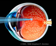
Complications of Laser Photocoagulations
Last updated: September 2026
Panretinal photocoagulation (PRP) and focal/grid laser remain mainstays of treating ischemic and edematous retinal disease — but the very mechanism that makes them effective (thermal tissue destruction) is also the source of every complication below. Newer laser delivery technology has meaningfully reduced, though not eliminated, these risks.
Common complications
- Visual field constriction — peripheral photoreceptor destruction from PRP predictably reduces peripheral visual field; relevant for driving assessment and patient counseling before treatment
- Reduced night vision (dark adaptation) — rod photoreceptors are disproportionately affected by peripheral scatter laser
- Macular edema — the single most common cause of visual acuity loss following PRP in classic series (accounting for roughly 8% of eyes with ≥2-line vision loss); inflammatory cytokine release from laser-induced tissue damage can exacerbate or trigger edema
- Choroidal effusions and exudative retinal detachment — more likely with high laser power/duration and dense treatment in a single sitting
- Transient angle closure and IOP spikes — from forward shift of the lens-iris diaphragm due to posterior segment swelling following heavy treatment
- Inadvertent foveal burns — a preventable but serious complication requiring meticulous attention to fixation and spot placement, especially near the fovea
- Progressive laser scar expansion over time, and rarely choroidal neovascularization arising from laser scars
Factors that increase complication risk
- High laser power and long pulse duration
- Dense treatment completed in a single session rather than split over multiple sittings
- Larger spot size and higher total burn count per session
Current mitigation strategies
- Multispot pattern lasers — deliver shorter-duration burns in a preset pattern, substantially reducing total thermal energy and treatment time compared to conventional single-spot argon lasers, while maintaining efficacy
- Subthreshold micropulse laser — delivers energy in very short pulses below the threshold for visible tissue damage, aiming to achieve therapeutic biological effect (particularly for macular edema) with minimal-to-no visible scarring — an active area of ongoing study for reducing laser-induced damage while preserving efficacy
- Splitting PRP into multiple sessions rather than one dense sitting, to reduce the risk of exudative detachment and angle closure
Key references: Panretinal Photocoagulation: A Review of Complications. Semin Ophthalmol. 2018;33(1). | Is Effective Photocoagulation Without Laser-Induced Damage Possible? Retina Today.
📘 Want the exam-ready deep dive? This topic, and much more is covered in Ophthalmology Explorer (A High-Yield Clinical Reference for Ophthalmology Residents, Fellows & Board Exams) — available on Kindle. See all my books.
Want to know more about treatment? Read about Retina Treatment at Sadbhaav or book a consultation with Dr. Dhaval Patel.