Layers in Ophthalmology
Cornea
epithelium, Bowman's layer, stroma proper, Descemet's
membrane, and endothelium
Sclera
episclera, scleral stroma proper, and lamina fusca
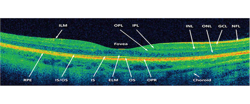
Retina
1. Retinal pigment epithelium
2. Photoreceptor cell layer
3. External limiting membrane
4. Outer nuclear layer
5. Outer plexiform layer
6. Inner nuclear layer
7. Inner plexiform layer
8. Ganglion cell layer
9. Nerve fiber layer
10. Internal limiting membrane
Tear Film
mucinous, aqueous, and lipid layers
The tear film is an exceedingly complex mixture of
secretions from multiple tissues and epithelia (Fig. 15.1) and consists of four
layers (Fig. 15.2). The innermost layer is a glycocalyx that extends from the
superficial layer of the ocular surface epithelia. The second is a mucous layer
that covers the glycocalyx and may mix with the third aqueous layer. The
outermost layer contains lipids. Similarly to mucous and aqueous layers,
aqueous and lipid layers may mix. Production and function of tear film layers
are distinct and will be presented separately.
Extra-ocular Muscles
an outer orbital layer composed of myofibers of extremely
small cross-sectional area and an inner global layer with myofibers larger than
in the orbital layer but still extremely small compared to non-cranial skeletal
muscle
Bruch's membrane
1) interrupted basement membrane of the
choriocapillaris, (2) outer collagenous zone, (3) elastic layer, (4) inner
collagenous zone, and (5) basement membrane of the RPE
Choroid
suprachoroid lamina (lamina fusca)
choroidal stroma
choriocapillaris
bruch’s membrane (basal lamina)
(The choroid is composed of the choriocapillaris layer, the
medium vessels layer (Sattler's layer) and the outer layer of large
vessels (Haller's
layer).)
Trabecular meshwork
uveal, corneoscleral, and juxtacanalicular.
Meninges
the dura mater, arachnoid layer, and pia mater.
Facial mimetic muscles (4)
A superficial layer consists of the orbicularis oculi,
zygomatic minor, and depressor anguli oris. The second layer contains the
platysma, depressor labii inferioris, the levator labii superioris alaeque
nasi, and zygomatic major muscles. The third layer consists of the levator
labii superioris and orbicularis oris. Finally, the deepest layer consists of
the mentalis, the levator anguli oris, and the buccinator. Branches of the
facial nerve supply the first three layers of muscles from the underside, while
the deepest layer is innervated from the outer surface.
Iris
Iris layers are described in many ways as per different references: I am quoting all here -
The Iris can be divided into four layers: (1) the anterior border layer, (2) stroma and sphincter muscle, (3) ante-rior epithelium and dilator muscle, and (4) posterior epithelium.
The Iris can be divided into four layers: (1) the anterior border layer, (2) stroma and sphincter muscle, (3) ante-rior epithelium and dilator muscle, and (4) posterior epithelium.
The iris is composed of five layers: the anterior border
layer, the iris stroma, the muscular layer, the anterior pigment epithelium,
and the posterior pigment epithelium. The clump cells of Koganei are part of
the iris stroma. The dilator and sphincter muscles make up the muscular layer
of the iris, which is anterior to the iris pigment epithelium. The color of the
iris is determined by the number and size of melanin pigment granules of the
stromal melanocytes.
The iris can be divided into two main layers: the posterior
leaf and the anterior leaf. The posterior iris leaf contains the
dilator muscle, the sphincter muscle, and the posterior pigmented epithelium.
From a front view of the iris, the dilator muscle is located circumferentially,
in the midperiphery of the iris.
Lateral geniculate nucleus
The primate lateral geniculate nucleus has six layers:
layers 1 and 2 consist of large (magnocellular) cells, and layers 3, 4, 5, and
6 consist of small (parvocellular) cells.
- compiled & published by Dr Dhaval Patel MD AIIMS