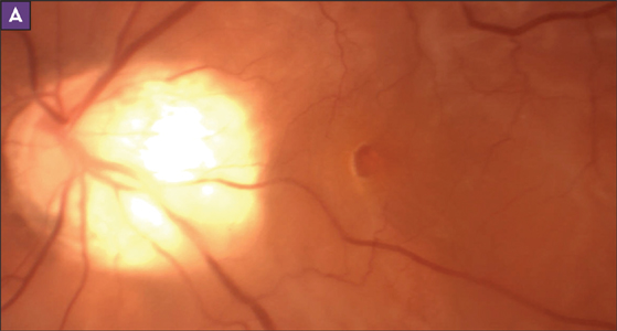
MHRD - Macular Hole Retinal Detachment
Last updated: September 2026
Macular hole retinal detachment (MHRD) is a distinct clinical entity seen almost exclusively in eyes with high (pathologic) myopia and posterior staphyloma — the combination of a full-thickness macular hole and an overlying retinal detachment confined largely to the posterior pole.
Mechanism
In highly myopic eyes, progressive axial elongation and posterior staphyloma formation create persistent tangential and anteroposterior vitreoretinal traction (from the epiretinal membrane, residual vitreous cortex, and the internal limiting membrane itself). This traction weakens the adhesion between the neurosensory retina and RPE at the fovea, eventually causing a macular hole through which liquefied vitreous gains access to the subretinal space — producing detachment localized to the posterior pole rather than a typical rhegmatogenous pattern.
Current first-line surgical management
Pars plana vitrectomy (PPV) combined with internal limiting membrane (ILM) peeling and tamponade is the established primary approach, with strong evidence supporting a specific refinement for these particularly challenging eyes:
- Inverted ILM flap technique is now supported by robust comparative data as superior to complete ILM peeling for MHRD in high myopia — head-to-head studies show significantly higher type 1 (complete) macular hole closure rates with the inverted flap (up to 100% in some series) versus conventional complete peeling (as low as 24% type 1 closure in comparative series), even though overall retinal reattachment rates are similar between the two techniques
- ICG or brilliant blue G staining is used to improve visualization during ILM peeling/flap creation
- Tamponade choice varies — gas (SF6/C3F8) or silicone oil depending on staphyloma severity and surgeon preference; silicone oil is often favored in eyes with extreme axial length or expected prolonged tamponade needs
Macular buckling: a resurging adjunct
For eyes with extreme myopia (axial length >30mm) and pronounced posterior staphyloma, adding a macular buckle to PPV can produce better anatomic and functional outcomes than PPV alone — by directly counteracting the posterior staphyloma's outward traction rather than only addressing the vitreous side of the traction. This is a useful option to be aware of for recurrent or particularly severe cases, though commercially available exoplants remain limited outside a few specific devices used mainly in Europe.
Emerging options for refractory cases
- Autologous retinal grafts and human amniotic membrane patching for eyes with limited ILM available for a flap
- Lens capsule flap transplantation is being studied as an alternative to ILM flap, offering a thicker and more manageable tissue
Key references: Inverted ILM flap technique for MHRD in high myopia vs. ILM peeling. 2025. | Prevalence and Management of Retinal Detachment in High Myopia. Retinal Physician. 2023. | Update on surgical management of complex macular holes: a review. 2021.
📘 Want the exam-ready deep dive? This topic, and much more is covered in Ophthalmology Explorer (A High-Yield Clinical Reference for Ophthalmology Residents, Fellows & Board Exams) — available on Kindle. See all my books.
Want to know more about treatment? Read about Retina Treatment at Sadbhaav or book a consultation with Dr. Dhaval Patel.