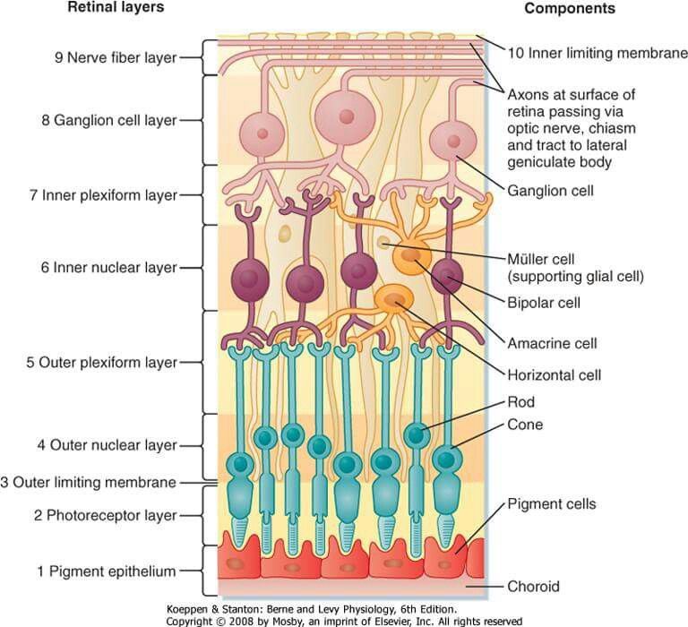
Retinal Layers: The 10 Layers and Which Layer Holds What
Last updated: October 2026
Key points in 60 seconds
- Ten layers from inside out: ILM, NFL, GCL, IPL, INL, OPL, ONL, ELM, photoreceptors, RPE.
- Flame haemorrhages and cotton-wool spots sit in the nerve fibre layer; dot and blot in the INL/OPL.
- Hard exudates and the macular star sit in the outer plexiform (Henle) layer; drusen lie between RPE and Bruch membrane.
- Müller cell nuclei are in the INL; photoreceptor nuclei in the ONL.
- The central retinal artery supplies the inner retina; the choroid supplies the outer retina, hence the cherry-red spot in CRAO.
Pages on this topic
- This page: 10 layers of the retina: quick table and which layer holds what
- Where lesions sit on OCT, layer by layer
- Layers of every eye structure, cornea to LGN
The ten layers of the retina, what each contains, and quick "which layer?" questions for revision. For the concept behind the layers, blood supply and OCT bands, see Retinal Layers & OCT Made Easy.
The 10 layers, from vitreous to choroid
| # | Layer | What it contains |
|---|---|---|
| 1 | Internal limiting membrane (ILM) | Müller cell footplates and their basement membrane |
| 2 | Nerve fibre layer (NFL) | Ganglion cell axons heading to the disc |
| 3 | Ganglion cell layer (GCL) | Ganglion cell bodies (and displaced amacrine cells) |
| 4 | Inner plexiform layer (IPL) | Synapses of bipolar and amacrine cells with ganglion cells |
| 5 | Inner nuclear layer (INL) | Nuclei of bipolar, horizontal, amacrine and Müller cells |
| 6 | Outer plexiform layer (OPL) | Synapses of photoreceptors with bipolar and horizontal cells (Henle fibre layer at the macula) |
| 7 | Outer nuclear layer (ONL) | Nuclei of rods and cones |
| 8 | External limiting membrane (ELM) | Junctions between Müller cells and photoreceptors |
| 9 | Photoreceptor layer | Rod and cone inner and outer segments |
| 10 | Retinal pigment epithelium (RPE) | Single pigmented cell layer on Bruch membrane |
Which layer? Quick revision
| Feature | Layer |
|---|---|
| Müller cell nuclei | Inner nuclear layer |
| Photoreceptor nuclei | Outer nuclear layer |
| Rod spherules and cone pedicles (synaptic triads) | Outer plexiform layer |
| Bipolar–amacrine–ganglion synapses | Inner plexiform layer |
| Cells projecting to the LGN and pretectum | Ganglion cell layer |
| Vitamin A storage and phagocytosis of outer segments | RPE |
| High dark current (cGMP-gated channels open in the dark) | Photoreceptor outer segments |
| Flame haemorrhages, cotton wool spots | Nerve fibre layer |
| Dot and blot haemorrhages | Inner nuclear / outer plexiform layer |
| Hard exudates, macular star | Outer plexiform (Henle) layer |
| Drusen | Between RPE and Bruch membrane |
Blood supply: the central retinal artery feeds the inner retina (NFL to INL); the choroid feeds the outer retina (ONL, photoreceptors, RPE). That is why CRAO spares the fovea's red colour.
Related: Retinal layers & OCT made easy, How to read an OCT, Layers in ophthalmology.
Revise with flip cards
📘 Want the exam-ready deep dive? This topic is covered in iNotes 2027: Basic Science & Optics (Ophthalmology Postgraduate Exam Notes) — available on Kindle. See all my books.
Experiencing symptoms like these? Learn about our Eye Department or book a consultation with Dr. Dhaval Patel.