Role of OCT Angiography in Detecting Early Diabetic Retinopathy
What is the role of OCT ANGIOGRAPHY in detecting Early Diabetic Retinopathy ?
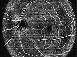
OCT angiography (OCTA) has emerged as a transformative tool in detecting diabetic retinopathy (DR) at stages where conventional methods may miss critical changes.
---What is OCTA?
OCT Angiography is a non-invasive imaging technique that maps retinal and choroidal vasculature by detecting motion contrast from flowing blood cells — without the need for dye injection (unlike Fluorescein Angiography/FFA).
---Why Early Detection Matters
Diabetic retinopathy often causes **irreversible vision loss before symptoms appear**. The window for effective intervention is in the **preclinical and early clinical stages** — exactly where OCTA excels.
---Key Roles of OCTA in Early DR Detection
1. Foveal Avascular Zone (FAZ) Analysis
2. Detection of Subclinical Capillary Changes
3. Retinal Flow Density Mapping
4. Layer-by-Layer Vascular Analysis
5. Choriocapillaris Assessment
6. Monitoring Diabetic Macular Ischemia (DMI)
| Feature | Fundus Photography | FFA | OCTA |
|---|---|---|---|
| Dye injection needed | No | Yes | No |
| Deep plexus imaging | Poor | Limited | Excellent |
| FAZ quantification | No | Qualitative | Quantitative |
| Subclinical ischemia | No | Limited | Yes |
| Microaneurysm detection | Limited | Good | Good–Excellent |
| Dynamic leakage info | No | Yes | No |
| Repeatability | High | Moderate | High |
Limitations to keep in mind
###| OCTA Limitation | Description & Clinical Impact |
|---|---|
| No leakage information | Cannot replace FFA entirely for assessing macular edema leakage. |
| Motion artifacts | Patient cooperation is essential to avoid image distortion. |
| Limited field of view | Standard coverage is only 3×3 or 6×6 mm, though wide-field OCTA is emerging. |
| Cost and availability | The technology is expensive and not universally accessible yet. |
| Interpretation learning curve | Requires specially trained readers to correctly analyze data. |
Clinical Significance Summary
OCTA enables detection of DR at the **neurovascular unit level**, identifying capillary dropout, FAZ changes, and perfusion deficits **before** lesions appear on fundoscopy — offering a critical opportunity for early intervention, tighter glycemic control counseling, and prevention of vision-threatening progression.
It is increasingly recommended as an adjunct in screening high-risk diabetic patients, particularly those with long disease duration, poor glycemic control, or subclinical visual symptoms.
These all details in just for basic information about the diseases. Please consult your doctor before following this by yourself. --- Dr Dhaval Patel (MD, AIIMS Delhi)
- compiled & published by Dr Dhaval Patel MD AIIMS