Ocular Physiology In Depth: Tear Film, Cornea, Aqueous, Lens, Retina and Vision
Last updated: November 2026
Physiology explains why diseases look the way they do and how our drugs and operations work. This in-depth guide covers each part of the eye in turn, from the tear film to the visual cortex, with the mechanisms and numbers that examiners ask.
Tear film
| Component | Source | Function |
|---|---|---|
| Lipid layer | Meibomian glands (and Zeis) | Reduces evaporation, stabilises the film |
| Aqueous-mucin gel | Main and accessory lacrimal glands (Krause, Wolfring); goblet cells | Nutrition, washing, antimicrobial proteins (lysozyme, lactoferrin, lipocalin, secretory IgA) |
| Glycocalyx | Membrane-bound mucins (MUC1, MUC4, MUC16) of the epithelium | Makes the surface wettable |
- Tear volume about 7 µL; secretion about 1.2 µL/min; turnover about 16% per minute.
- Reflex secretion: afferent V1, efferent parasympathetic fibres from the superior salivatory nucleus via CN VII, the greater petrosal nerve and the pterygopalatine ganglion.
- Tear osmolarity normally about 300 mOsm/L; above about 308 or an inter-eye difference of more than 8 suggests dry eye.
- Drainage depends on the lacrimal pump: blinking compresses the canaliculi and sac.
Cornea
- Transparency: stromal collagen fibrils of uniform diameter, spaced less than half a wavelength apart, so scattered light cancels out (Maurice's lattice theory); no vessels; non-keratinised epithelium; and relative dehydration.
- Deturgescence (about 78% water) is kept by the endothelial pump (Na+/K+-ATPase and bicarbonate transport) and the barrier function of the epithelium and endothelium. Stromal swelling pressure is about 50–60 mmHg.
- The endothelium does not divide; cells spread to cover gaps. Decompensation occurs below about 500 cells/mm².
- Oxygen comes mostly from the tear film (air) with eyes open and from the palpebral conjunctiva with eyes closed: why overnight contact lens wear causes hypoxia.
- Epithelial wound healing: cells slide in hours, proliferate and stratify over days; Bowman layer does not regenerate.
Aqueous humour and IOP
| Process | Details |
|---|---|
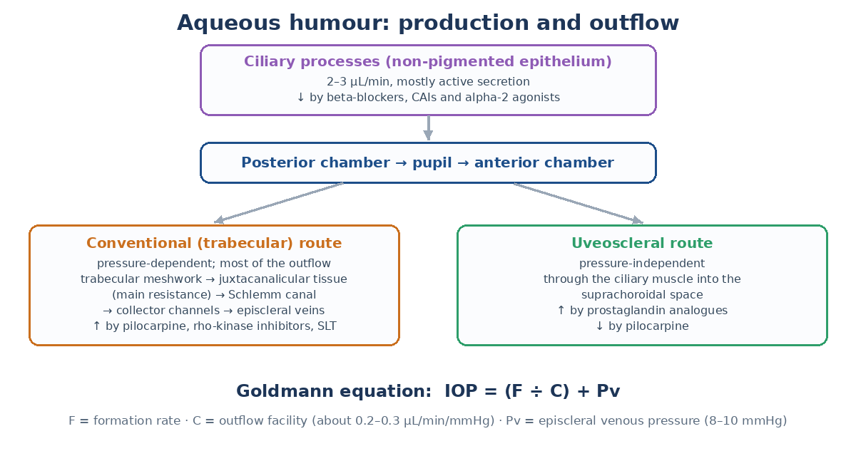
| Production | About 2–3 µL/min by the non-pigmented ciliary epithelium: active secretion (the major part; needs Na+/K+-ATPase and carbonic anhydrase), ultrafiltration and diffusion. Lower at night. |
| Composition | Low protein (about 1/200 of plasma), high ascorbate (about 15–20 times plasma, protects against UV and oxidation), lactate; glucose slightly below plasma |
| Conventional outflow | Trabecular meshwork to Schlemm canal: pressure-dependent, most of the outflow; resistance mainly in the juxtacanalicular tissue |
| Uveoscleral outflow | Through the ciliary muscle into the suprachoroidal space: pressure-independent; increased by prostaglandin analogues, reduced by pilocarpine |
| Outflow facility | About 0.2–0.3 µL/min/mmHg; falls with age and in POAG |
| Episcleral venous pressure | About 8–10 mmHg; raised in carotid-cavernous fistula and Sturge-Weber syndrome |
Goldmann equation: IOP = (F ÷ C) + Pv. IOP is normally about 10–21 mmHg, with a diurnal variation of 3–6 mmHg (often highest in the early morning, partly when lying down). Ocular perfusion pressure = blood pressure − IOP.
Blood-ocular barriers
- Blood-aqueous barrier: tight junctions of the non-pigmented ciliary epithelium and the iris vessel endothelium; breaks down in uveitis and after surgery (flare).
- Inner blood-retina barrier: tight junctions of retinal capillary endothelium (breaks down in diabetic macular oedema and vein occlusion).
- Outer blood-retina barrier: tight junctions of the RPE (breaks down in CSCR, choroidal neovascularisation).
- Fenestrated choroidal capillaries leak fluorescein freely; retinal vessels do not, which is why FFA shows retinal leakage clearly.
Lens
- Avascular and anucleate in its core; energy mainly from anaerobic glycolysis (about 80% of glucose), plus the hexose monophosphate shunt for NADPH.
- Antioxidant defences: glutathione (very high concentration), ascorbate, superoxide dismutase.
- Transparency: tightly packed fibres with few organelles and crystallin proteins (alpha, beta, gamma) at high concentration (about 33% of lens weight, the highest protein content of any tissue). Alpha-crystallin acts as a chaperone.
- Ion balance by Na+/K+-ATPase in the epithelium (pump-leak system).
- Cataract mechanisms: oxidation and protein cross-linking (age, UV), osmotic swelling from sorbitol via aldose reductase (diabetes, galactosaemia), steroids (posterior subcapsular), and hydration changes.
Vitreous
- A gel of type II collagen fibrils kept apart by hyaluronan; liquefies with age (synchysis) and collapses (syneresis), leading to posterior vitreous detachment, usually after age 50 (earlier in myopes).
- It acts as a metabolic reservoir and a barrier to diffusion and cell migration; vitrectomy speeds lens oxidation (nuclear cataract) and drug clearance.
Retina and RPE
| RPE function | Clinical link |
|---|---|
| Absorbs stray light (melanin) | Albinism: glare, poor acuity |
| Phagocytoses shed outer segment discs (about 10% daily) | Failure leads to debris and degeneration (some RP genes) |
| Recycles retinal (visual cycle: RPE65) | RPE65 Leber amaurosis treated with gene therapy |
| Pumps fluid from subretinal space to choroid | Subretinal fluid in CSCR when the pump or barrier fails |
| Forms the outer blood-retina barrier | Leakage on FFA |
| Secretes growth factors (VEGF towards choroid, PEDF towards retina) | Basis of choroidal neovascularisation and anti-VEGF therapy |
- Müller cells span the retina: structural support, potassium and glutamate buffering, and they contribute to the ERG b-wave.
- Macular pigment (lutein, zeaxanthin) in the Henle fibre and inner layers filters blue light (the dark spot on FAF).
- The retina has one of the highest oxygen consumptions in the body; the outer retina is supplied by the choroid, the inner retina by the retinal circulation.
Phototransduction
- A photon isomerises 11-cis-retinal to all-trans-retinal in rhodopsin (peak absorption about 498 nm).
- Activated rhodopsin activates transducin (a G protein), which activates phosphodiesterase.
- cGMP falls, cGMP-gated sodium channels close, and the "dark current" stops.
- The photoreceptor hyperpolarises and releases less glutamate; bipolar cells respond (ON cells depolarise, OFF cells hyperpolarise).
- Recovery: rhodopsin kinase and arrestin stop the cascade; calcium-dependent feedback restores cGMP.
All-trans-retinal is carried to the RPE, converted back to 11-cis-retinal (RPE65 isomerase) and returned: the visual cycle. Cones also use a Müller cell pathway for faster regeneration.
Adaptation and colour vision
- Dark adaptation: a cone phase (best in 5–10 minutes), the rod-cone break at about 7–10 minutes, then the rod phase reaching maximum sensitivity at about 30–40 minutes. Delayed in vitamin A deficiency, RP and AMD.
- Light adaptation is faster (seconds to minutes).
- Photopic vision uses cones (peak sensitivity 555 nm), scotopic vision uses rods (507 nm); the shift is the Purkinje shift.
- Trichromacy: S cones (about 420 nm), M cones (530 nm) and L cones (560 nm); then opponent processing (red-green, blue-yellow, light-dark) in the retina and LGN.
- Congenital red-green defects are X-linked (about 8% of men); acquired optic nerve disease tends to cause red-green defects and retinal disease blue-yellow (Kollner rule).
Pupil and accommodation
- Light reflex: retina, optic nerve and tract (pupillary fibres leave before the LGN), pretectal nuclei (bilateral, giving the consensual response), Edinger-Westphal nuclei, CN III, ciliary ganglion, short ciliary nerves, sphincter.
- Near triad: accommodation, convergence and miosis; the near pathway runs more ventrally, explaining light-near dissociation.
- Accommodation (Helmholtz): the ciliary muscle contracts, zonular tension falls, and the lens rounds up, especially its anterior surface. Amplitude falls from about 14 D at age 10 to about 1 D at age 60 (presbyopia).
- Sympathetic dilation via the three-neuron pathway; the pupil is the balance of sphincter and dilator tone.
Ocular blood flow
- The retinal circulation is autoregulated (no autonomic innervation) and has a high oxygen extraction; the choroidal circulation has very high flow, low extraction and sympathetic control, and is poorly autoregulated.
- Failure of autoregulation and low perfusion pressure are proposed in normal-tension glaucoma.
- Fluorescein reaches the choroid about 1 second before the retinal arteries (arm-retina time about 10–15 seconds).
Visual function
- Visual acuity is limited by foveal cone spacing (about 0.5 minute of arc) and optics; 6/6 corresponds to resolving 1 minute of arc.
- Contrast sensitivity peaks at about 3–5 cycles per degree.
- Binocular single vision needs normal retinal correspondence and fusion within Panum fusional area; stereopsis arises from horizontal disparity.
- Electrophysiology basis: ERG a-wave from photoreceptors, b-wave from bipolar and Müller cells, oscillatory potentials from amacrine cells; EOG from the RPE (light rise / dark trough, the Arden ratio); VEP from the visual cortex. See electrophysiology made easy.
Where residents slip
- Saying photoreceptors depolarise to light.
- Thinking prostaglandins act on the trabecular route (mainly uveoscleral).
- Forgetting that the endothelium does not regenerate.
- Confusing the photopic (555 nm) and scotopic (507 nm) peaks.
- Saying the retinal circulation has autonomic control (it is autoregulated).
Test yourself: flip cards
Quick quiz
Related: Ocular Anatomy In Depth, ERG, EOG and VEP Made Easy: One Test per Level, Ocular Pharmacology In Depth, Revise ophthalmology checklist.
Key sources
- Levin LA, et al. Adler's Physiology of the Eye. 11th ed. Elsevier; 2011
- American Academy of Ophthalmology. BCSC Section 2: Fundamentals and Principles of Ophthalmology (current edition)
- Forrester JV, et al. The Eye: Basic Sciences in Practice. 5th ed. Elsevier; 2020
- Willcox MDP, et al. TFOS DEWS II Tear Film Report. Ocul Surf. 2017;15:366-403
📘 Want more exam-ready material? See Most Commons in Ophthalmology on Kindle, or browse all my books.
Looking for an eye specialist in Ahmedabad? Learn about our Eye Department or book a consultation with Dr. Dhaval Patel.