Ocular Anatomy In Depth: Globe, Angle, Retina, Optic Nerve and Blood Supply
Last updated: November 2026
Clinical ophthalmology is applied anatomy: the angle explains glaucoma, the blood supply explains vascular occlusions, and the layers of the retina explain every OCT. This guide goes structure by structure, with the measurements and relations that examiners ask and surgeons use.
The globe
| Measurement | Typical adult value |
|---|---|
| Axial length | About 23.5–24 mm (16–17 mm at birth) |
| Volume | About 6.5–7 mL |
| Coats | Outer fibrous (cornea, sclera), middle vascular (uvea), inner neural (retina) |
| Chambers | Anterior chamber about 0.25 mL, depth about 3 mm centrally; posterior chamber about 0.06 mL; vitreous cavity about 4 mL |
Cornea
| Feature | Value or fact |
|---|---|
| Diameter | About 11.5–12 mm horizontally, 10.5–11 mm vertically (horizontally oval from the front) |
| Thickness | About 540 µm centrally, 650–700 µm peripherally |
| Radius of curvature | Anterior about 7.8 mm, posterior about 6.5 mm |
| Power | About +43 D in total (anterior surface about +49 D, posterior about −6 D): two-thirds of the eye's focusing power |
| Nerve supply | Long ciliary nerves from the nasociliary branch of V1; the densest sensory innervation in the body |
| Layer | Key facts |
|---|---|
| Epithelium | Non-keratinised stratified squamous, 5–7 cell layers, about 50 µm; renews in about 7 days from limbal stem cells |
| Bowman layer | Acellular condensed collagen, 8–12 µm; does not regenerate (scars after injury) |
| Stroma | About 90% of thickness; 200–250 lamellae of regularly spaced type I collagen with keratocytes |
| Pre-Descemet (Dua) layer | A thin, tough acellular layer (about 10–15 µm) described in 2013; relevant to big-bubble DALK |
| Descemet membrane | Basement membrane of the endothelium; thickens with age (about 3 µm at birth to 10 µm or more); site of guttae in Fuchs dystrophy |
| Endothelium | Single layer of hexagonal cells; about 2,500–3,000 cells/mm² in young adults, falling with age; does not divide in humans |
Sclera and limbus
- Sclera: mainly type I collagen; thickest posteriorly (about 1 mm), thinnest just behind the rectus insertions (about 0.3 mm), the usual site of rupture in blunt trauma.
- The lamina cribrosa is the sieve-like posterior scleral opening for the optic nerve axons.
- The limbus is a 1–2 mm transition zone; limbal epithelial stem cells live in the palisades of Vogt. The surgical limbus is the landmark for incisions.
- Rectus insertions follow the spiral of Tillaux: medial about 5.5 mm, inferior 6.5 mm, lateral 6.9 mm, superior 7.7 mm from the limbus.
The anterior chamber angle
| Structure (from front to back on gonioscopy) | Notes |
|---|---|
| Schwalbe line | The end of Descemet membrane: a fine white line, sometimes pigmented (Sampaolesi line in pseudoexfoliation) |
| Trabecular meshwork | Uveal (innermost), corneoscleral, and juxtacanalicular meshwork (the main site of outflow resistance); the pigmented part overlies Schlemm canal |
| Scleral spur | A white band; the attachment of the longitudinal ciliary muscle |
| Ciliary body band | Grey-brown; wide in myopes and after angle recession |
| Iris root | Insertion of the iris into the ciliary body |
Aqueous leaves through Schlemm canal, collector channels and aqueous veins into the episcleral veins (conventional route), or through the ciliary muscle into the suprachoroidal space (uveoscleral route).
Uvea
| Part | Key anatomy |
|---|---|
| Iris | Anterior border layer, stroma, sphincter pupillae (parasympathetic, CN III via the ciliary ganglion and short ciliary nerves), dilator pupillae (sympathetic, via long ciliary nerves), and a two-layered posterior pigment epithelium |
| Ciliary body | About 6 mm wide: pars plicata (anterior 2 mm, about 70 ciliary processes that make aqueous) and pars plana (posterior 4 mm, the safe entry zone for vitrectomy and injections). Ciliary muscle has longitudinal, radial and circular fibres |
| Choroid | Haller layer (large vessels), Sattler layer (medium), choriocapillaris (fenestrated, the highest blood flow per gram of any tissue); thickest at the posterior pole |
| Bruch membrane | Five layers: RPE basement membrane, inner collagenous, elastic, outer collagenous, choriocapillaris basement membrane; deposits here underlie drusen and AMD |
Lens
- About 9–10 mm in diameter and 4 mm thick, growing thicker throughout life; refractive power about +20 D in the eye.
- Capsule: the thickest basement membrane in the body; thickest at the anterior pre-equatorial zone (about 14 µm), thinnest at the posterior pole (about 4 µm), which is why the posterior capsule ruptures easily.
- Epithelium only on the anterior surface; equatorial cells form new fibres throughout life, compressing old fibres into the nucleus.
- Sutures: an upright Y anteriorly and an inverted Y posteriorly.
- Zonules arise from the pars plana and pars plicata and insert around the equator.
Vitreous
- About 4 mL, 99% water, with type II collagen fibrils and hyaluronan.
- Firmest attachment at the vitreous base, which straddles the ora serrata (about 2 mm in front and 3 mm behind); also at the disc, the fovea, retinal vessels and the posterior lens capsule (Wieger ligament, enclosing Berger space).
- Cloquet canal is the remnant of the hyaloid artery path; Mittendorf dot and Bergmeister papilla are its remnants.
Retina
| Layer (inner to outer) | Contains or corresponds to |
|---|---|
| Internal limiting membrane | Basement membrane of Müller cells; peeled in macular hole surgery |
| Nerve fibre layer | Ganglion cell axons; flame haemorrhages, cotton wool spots |
| Ganglion cell layer | Ganglion cell bodies; thickest around the fovea |
| Inner plexiform layer | Synapses of bipolar, amacrine and ganglion cells |
| Inner nuclear layer | Bipolar, horizontal, amacrine and Müller cell nuclei; dot-blot haemorrhages, cysts |
| Outer plexiform layer | Photoreceptor-bipolar synapses; Henle fibre layer at the fovea (macular star, hard exudates) |
| Outer nuclear layer | Photoreceptor nuclei |
| External limiting membrane | Junctions between Müller cells and photoreceptors |
| Photoreceptor layer | Inner and outer segments; the ellipsoid zone on OCT |
| Retinal pigment epithelium | Single layer of hexagonal cells resting on Bruch membrane |
- Macula about 5.5 mm across; fovea about 1.5 mm; foveola about 0.35 mm; foveal avascular zone about 0.5 mm. The foveola contains only cones.
- About 120 million rods and 6 million cones; cone density peaks at the foveola, rod density about 20° from fixation.
- Ora serrata: the anterior end of the retina, about 6 mm (nasal) to 7 mm (temporal) behind the limbus.
Optic nerve
| Segment | Length |
|---|---|
| Intraocular (disc) | About 1 mm |
| Intraorbital | 25–30 mm (slack, allowing eye movement) |
| Intracanalicular | 6–10 mm (vulnerable in trauma) |
| Intracranial | About 10 mm, to the chiasm |
- About 1.2 million ganglion cell axons; the disc is about 1.5–1.8 mm across, slightly taller than wide.
- The orbital and intracranial nerve is wrapped in dura, arachnoid and pia: raised intracranial pressure is transmitted along the sheath (papilloedema).
- Blood supply of the disc: the surface nerve fibre layer from the central retinal artery; the prelaminar and laminar parts from the short posterior ciliary arteries (circle of Zinn-Haller): the territory affected in NAION and GCA.
Blood supply
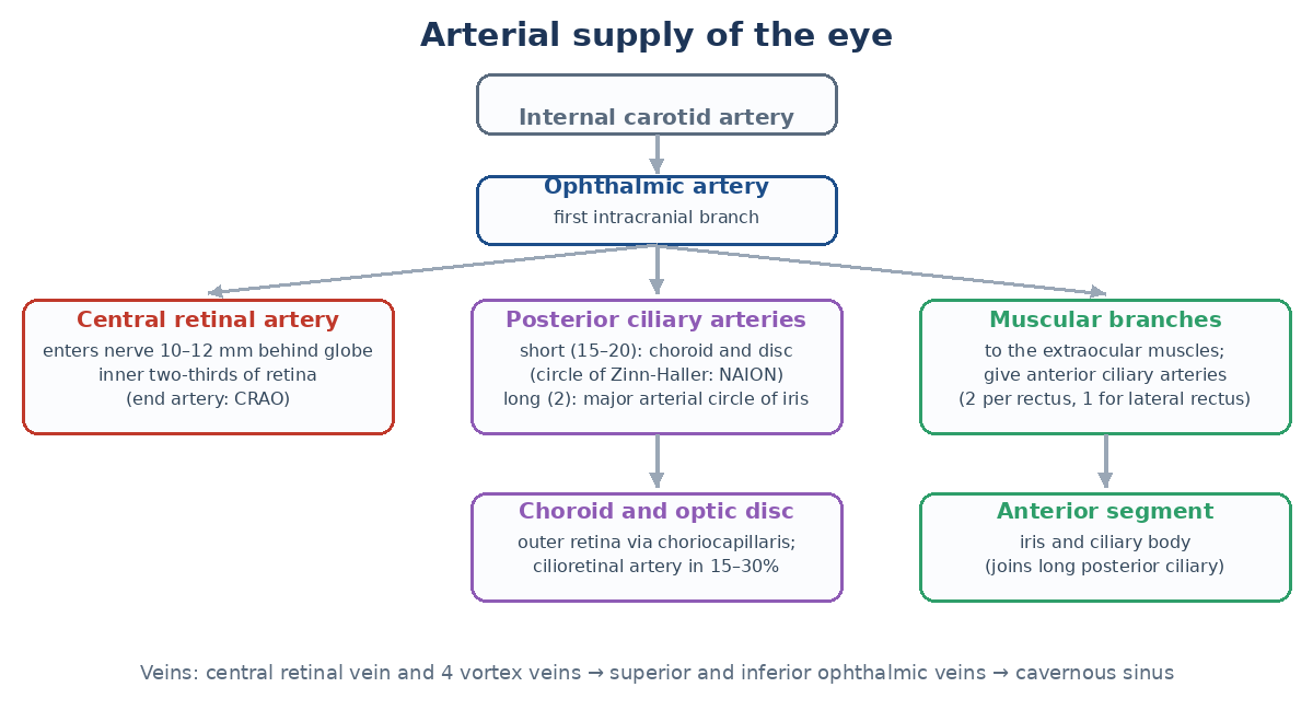
- The ophthalmic artery is the first intracranial branch of the internal carotid artery, entering the orbit through the optic canal below the nerve.
- Central retinal artery: enters the optic nerve about 10–12 mm behind the globe and supplies the inner two-thirds of the retina (an end artery: occlusion causes a cherry-red spot).
- Posterior ciliary arteries: short (about 15–20 branches) to the choroid and optic nerve head; two long ones run in the horizontal meridian to the major arterial circle of the iris.
- Anterior ciliary arteries: from the muscular branches, two per rectus muscle except the lateral rectus (one), so operating on many recti at once risks anterior segment ischaemia.
- A cilioretinal artery (from the choroidal circulation) is present in about 15–30% of eyes and can spare central vision in CRAO.
- Veins: the central retinal vein and the vortex veins (usually four, one per quadrant) drain into the superior and inferior ophthalmic veins and then the cavernous sinus.
Nerves
| Nerve or ganglion | Role |
|---|---|
| Ciliary ganglion | In the orbit lateral to the optic nerve; the parasympathetic root (from the CN III inferior division, via the nerve to inferior oblique) synapses here; sensory and sympathetic fibres pass through without synapsing |
| Short ciliary nerves | Carry parasympathetic fibres to the sphincter and ciliary muscle, plus sensory and sympathetic fibres |
| Long ciliary nerves | Sensory to the cornea; sympathetic to the dilator pupillae |
| CN III, IV, VI | Oculomotor (all recti except lateral, inferior oblique, levator, pupil), trochlear (superior oblique), abducens (lateral rectus) |
| CN VII | Orbicularis oculi (closure) and reflex tear secretion via the pterygopalatine ganglion |
| Sympathetic chain | Three-neuron pathway (hypothalamus, ciliospinal centre of Budge at C8–T2, superior cervical ganglion) to the dilator and Müller muscle: interrupted in Horner syndrome |
Lids and lacrimal system
- Lid layers: skin, orbicularis, orbital septum, preaponeurotic fat, levator aponeurosis and Müller muscle (upper lid), tarsus and conjunctiva. The grey line (muscle of Riolan) divides anterior and posterior lamellae.
- Tarsal plates: upper about 10 mm high, lower about 4–5 mm; meibomian glands (about 25 upper, 20 lower).
- Lid retractors: levator (CN III), Müller muscle (sympathetic, 2 mm of lift), and the capsulopalpebral fascia in the lower lid.
- Lacrimal gland: orbital and palpebral lobes divided by the levator aponeurosis; ducts open into the superotemporal fornix.
- Drainage: puncta, canaliculi (2 mm vertical then 8 mm horizontal), common canaliculus, sac (valve of Rosenmüller), nasolacrimal duct (about 12–18 mm) opening into the inferior meatus under the valve of Hasner.
For the orbit, see orbital anatomy made easy; for development, embryology made easy; for histological layers, layers of the eye.
Where residents slip
- Saying the posterior capsule is the thickest part of the lens capsule (it is the thinnest).
- Forgetting the lateral rectus has only one anterior ciliary artery.
- Mixing up which artery supplies the disc in NAION (short posterior ciliary) versus the inner retina (central retinal artery).
- Placing pars plana entry too far forward: 3.5–4 mm behind the limbus is the safe zone.
Test yourself: flip cards
Quick quiz
Related: Orbital Anatomy Made Easy: Walls, Fissures and Muscles, Layers in Ophthalmology: Cornea to LGN in Tables, Ocular Physiology Basics: Tear Film, Aqueous, Cornea, Lens and Retina, Revise ophthalmology checklist.
Key sources
- American Academy of Ophthalmology. BCSC Section 2: Fundamentals and Principles of Ophthalmology (current edition)
- Snell RS, Lemp MA. Clinical Anatomy of the Eye. 2nd ed. Blackwell Science; 1998
- Remington LA. Clinical Anatomy and Physiology of the Visual System. 3rd ed. Elsevier; 2012
- Dua HS, et al. Human corneal anatomy redefined: a novel pre-Descemet layer (Dua's layer). Ophthalmology. 2013;120:1778-1785
📘 Want more exam-ready material? See Most Commons in Ophthalmology on Kindle, or browse all my books.
Looking for an eye specialist in Ahmedabad? Learn about our Eye Department or book a consultation with Dr. Dhaval Patel.