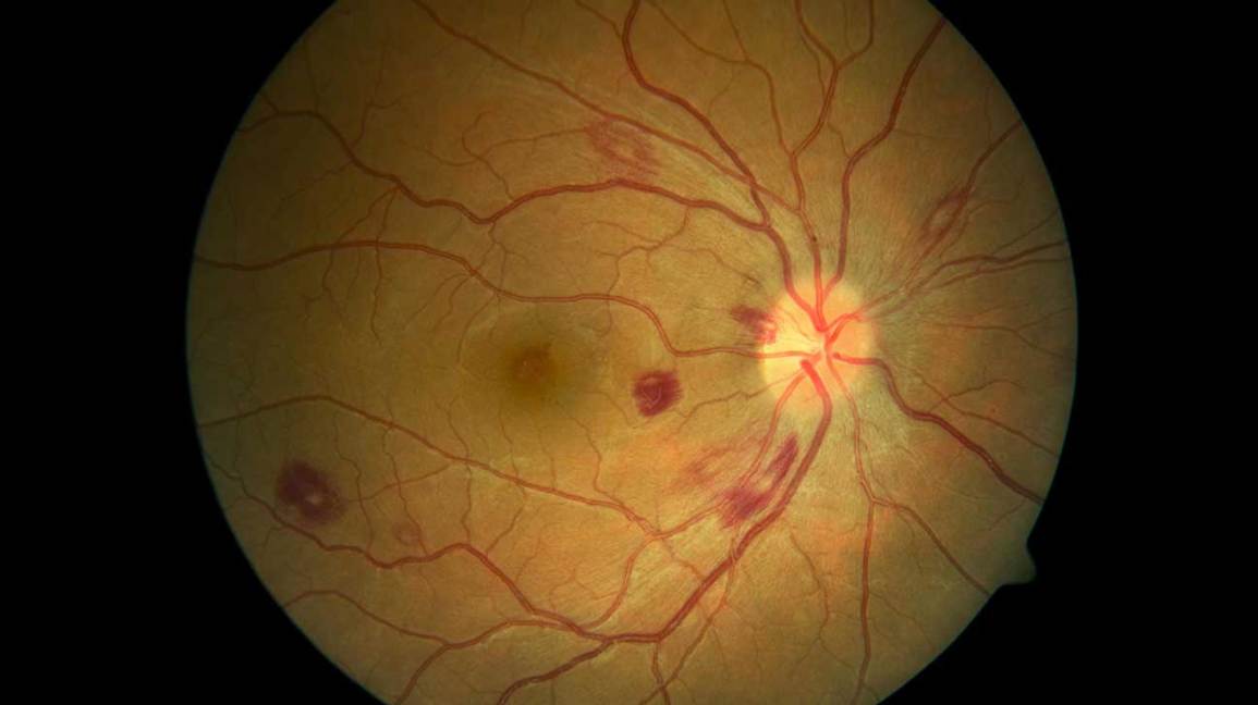
Dots & Spots in Ophthalmology: 19 Named Signs in Tables
Last updated: October 2026
Named dots and spots in ophthalmology, grouped by where you see them. For lines and rings (Hudson–Stähli, Fleischer, Vossius and more) see Lines & Rings.
Conjunctiva, cornea, iris and lens
| Name | What it is | Seen in |
|---|---|---|
| Bitot spot | Foamy white patch of keratinised conjunctiva, temporal to the limbus | Vitamin A deficiency (WHO X1B) |
| Horner–Trantas dots | White limbal collections of eosinophils and epithelial cells | Vernal keratoconjunctivitis |
| Koplik spots | White spots on the buccal mucosa (and rarely the conjunctiva/caruncle) | Measles |
| Krachmer spots (Kaye dots) | Subepithelial infiltrates in a graft | Corneal graft rejection |
| Cogan senile scleral plaque | Grey translucent hyaline patch just in front of a horizontal rectus insertion | Normal ageing |
| Brushfield spots | Grey-white spots in the peripheral iris | Down syndrome |
| Mittendorf dot | Small white dot on the posterior lens capsule (nasal to centre) | Remnant of the hyaloid artery |
Retina and choroid
| Name | What it is | Seen in |
|---|---|---|
| Cotton wool spots | Fluffy white nerve fibre layer infarcts | Diabetes (commonest), hypertension, HIV, lupus, emboli, carotid disease |
| Roth spots | White-centred retinal haemorrhages | Endocarditis, leukaemia, severe anaemia |
| Cherry-red spot | Red fovea surrounded by white retina | CRAO, Tay–Sachs, Niemann–Pick, Sandhoff, commotio retinae |
| Elschnig spots | Pale or pigmented patches over choroidal infarcts | Malignant hypertension, pre-eclampsia |
| Siegrist streaks | Linear pigment along choroidal vessels | Severe hypertension |
| Fuchs spot | Pigmented scar from regressed choroidal neovascularisation | Pathological myopia |
| Histo spots | Punched-out chorioretinal scars | Presumed ocular histoplasmosis syndrome |
| Gunn dots | Glistening reflexes from the internal limiting membrane near the disc | Normal young eyes |
| Birdshot spots | Cream ovoid spots radiating from the disc | Birdshot chorioretinopathy (HLA-A29) |
| Dalen–Fuchs nodules | Yellow-white sub-RPE nodules | Sympathetic ophthalmia, VKH, sarcoid |
| Leopard spots | Patchy hyper- and hypofluorescence on angiography from sub-RPE infiltrates | Vitreoretinal lymphoma, uveal effusion |
| Drusen | Yellow deposits between RPE and Bruch membrane | AMD (hard and soft), optic disc drusen are different |
Related: Lines and rings, Eponymous signs, Pathognomonic signs.
Revise with flip cards
Key sources
- Bowling B. Kanski's Clinical Ophthalmology, 9th ed. Elsevier; 2020
📘 Want the exam-ready deep dive? This topic, and much more is covered in Ophthalmology Explorer (A High-Yield Clinical Reference for Ophthalmology Residents, Fellows & Board Exams) — available on Kindle. See all my books.
Experiencing symptoms like these? Learn about our Eye Department or book a consultation with Dr. Dhaval Patel.