Approach to Diplopia: Monocular vs Binocular and the Three-Step Test
Last updated: November 2026
Double vision can come from a dry eye or an aneurysm. A structured history and four quick questions sort most patients in minutes and tell you who needs same-day imaging.
Four questions
| Question | Why it matters |
|---|---|
| 1. Does it go when one eye is covered? | No (persists with one eye open) = monocular: dry eye, cataract, irregular astigmatism, keratoconus, IOL edge, macular disease. Not neurological. |
| 2. Are the images side by side or one above the other? | Horizontal: VI nerve palsy, INO, convergence problems, decompensated phoria. Vertical: IV nerve palsy, skew deviation, TED, orbital floor fracture, myasthenia. |
| 3. Is it worse in one direction of gaze or at near or distance? | Points to the weak muscle; worse at distance suggests VI nerve palsy or divergence insufficiency. |
| 4. Does it vary through the day? | Fatigable, variable diplopia with ptosis: myasthenia gravis. |
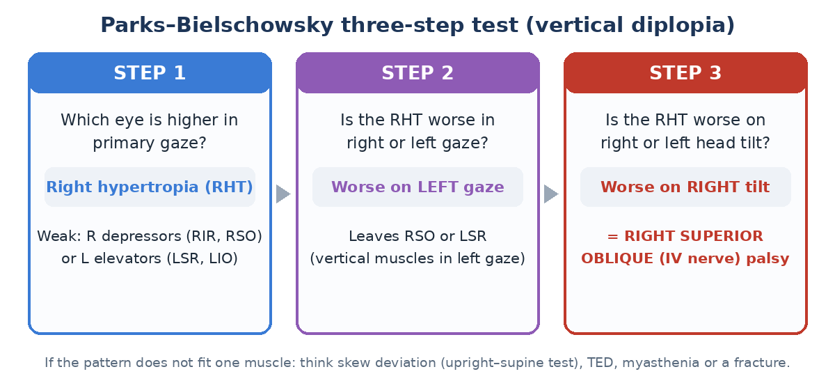
The three-step test (vertical diplopia)
- Step 1: which eye is higher in primary gaze?
- Step 2: is the hypertropia worse in right or left gaze?
- Step 3: is it worse with head tilt to the right or left (Bielschowsky)?
- Example: right hypertropia, worse on left gaze and right head tilt = right superior oblique (IV nerve) palsy. Check for a skew deviation (upright-supine test) if the pattern does not fit.
Red flags: image the same day
- A pupil-involving or painful III nerve palsy (aneurysm until proven otherwise: CT or MR angiography).
- Multiple cranial nerve palsies, or palsy with other neurological signs.
- Diplopia with headache and papilloedema.
- Age over 50 with jaw claudication or scalp tenderness: giant cell arteritis.
- A palsy in a young patient without vascular risk factors, or one that progresses or does not recover in 3 months.
Common causes and tests
| Cause | Clues | Tests |
|---|---|---|
| Microvascular (ischaemic) palsy | Older, diabetic or hypertensive, pupil-sparing III, isolated IV or VI; recovers in 3 months | BP, sugar, ESR and CRP |
| Myasthenia gravis | Variable, fatigable, ptosis, Cogan lid twitch | Ice-pack test, acetylcholine receptor and MuSK antibodies, single-fibre EMG, CT chest |
| Thyroid eye disease | Restriction, lid retraction, proptosis; inferior rectus most often | Thyroid tests, orbital imaging |
| Decompensated phoria | Long-standing tendency, comitant, no other signs | Prism cover test, old photographs |
Where residents slip
- Imaging every monocular diplopia.
- Missing myasthenia because the eye movements look normal at that moment.
- Calling a pupil-involving III nerve palsy microvascular.
- Not checking ESR and CRP in older patients.
Test yourself: flip cards
Related: Third, Fourth & Sixth Nerve Palsies, Ocular Motility Made Easy: Muscle Actions, Palsies, Three-Step, Internuclear Ophthalmoplegia (INO).
Key sources
- Kanski JJ, Bowling B. Clinical Ophthalmology: A Systematic Approach (strabismus and neuro-ophthalmology chapters)
- Parks MM. Isolated cyclovertical muscle palsy. Arch Ophthalmol. 1958;60:1027-1035
- Tamhankar MA, et al. Isolated third, fourth and sixth cranial nerve palsies from presumed microvascular versus other causes. Ophthalmology. 2013;120:2264-2269
📘 Want more exam-ready material? See Most Commons in Ophthalmology on Kindle, or browse all my books.
Looking for an eye specialist in Ahmedabad? Learn about our Eye Department or book a consultation with Dr. Dhaval Patel.