Third, Fourth & Sixth Nerve Palsies
Last updated: September 2026
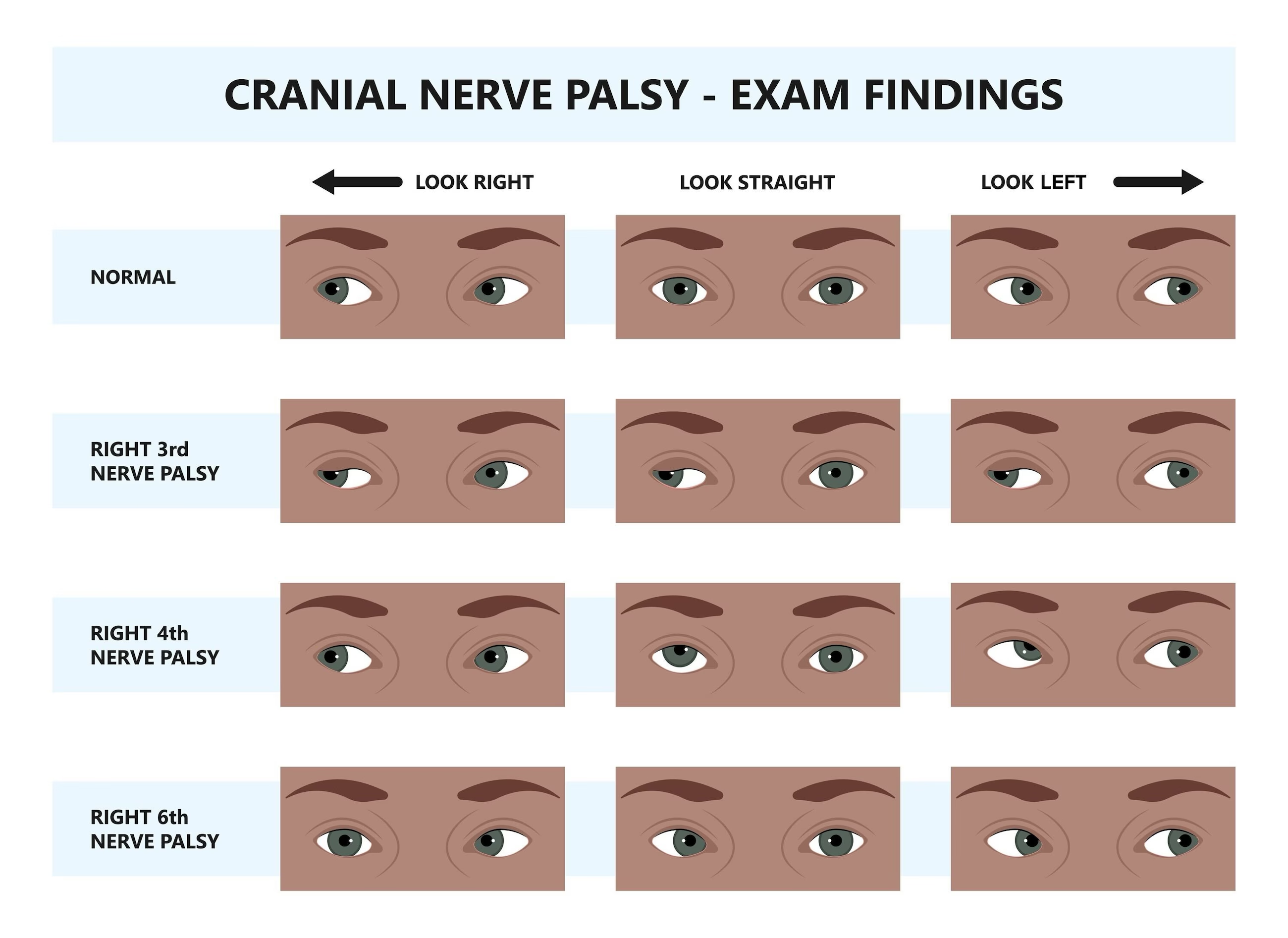
The third, fourth, and sixth cranial nerves each produce a distinct pattern of diplopia and misalignment — recognizing the pattern lets you localize before you even order imaging, and knowing which presentations are dangerous versus benign changes how urgently you act.
Third nerve (oculomotor) palsy
- Classic picture: ptosis, "down and out" eye position, and (if pupil-involving) a dilated, poorly reactive pupil
- The single most important question: is the pupil involved? Pupillary fibers run peripherally in the nerve, making them vulnerable to compression — a pupil-involving CN III palsy is a compressive lesion (most importantly, a posterior communicating artery aneurysm) until proven otherwise, and warrants urgent CTA/MRA
- A pupil-sparing complete CN III palsy in a patient with vascular risk factors (diabetes, hypertension) is more often microvascular/ischemic — but this is a diagnosis of exclusion, and any patient should be re-examined within days, since compressive lesions can evolve to involve the pupil over time
- Aberrant regeneration (eyelid retraction on downgaze, pupil constriction on adduction) suggests a chronic compressive or congenital process rather than an acute microvascular event, and should prompt imaging even in an older patient
Fourth nerve (trochlear) palsy
- The trochlear nerve has the longest intracranial course of any cranial nerve and is the thinnest — making it especially vulnerable to closed head trauma, even relatively minor trauma
- Classic picture: vertical diplopia, worse on downgaze and on gaze away from the affected side, with a compensatory head tilt away from the affected eye (patients often adopt this posture long before presenting)
- Three-step test (Parks-Bielschowsky) helps confirm and localize: identify the hypertropic eye, then whether it worsens on gaze to one side, then whether it worsens on head tilt to one side — worsening on head tilt toward the hypertropic eye implicates that eye's superior oblique
- Congenital fourth nerve palsies are common and often decompensate in adulthood — old photographs showing a long-standing head tilt are a genuinely useful clue that this isn't a new, acute problem
Sixth nerve (abducens) palsy
- Classic picture: horizontal diplopia, worse at distance, with an esotropia that increases on gaze toward the affected side (inability to abduct that eye)
- The abducens nerve has a long subarachnoid course, making it particularly susceptible to stretching from raised intracranial pressure — a sixth nerve palsy is a classic "false localizing sign," since it can result from generalized raised ICP rather than a lesion directly on the nerve's path
- In a recent hospital-based series, sixth nerve palsy was the second most common ocular cranial nerve palsy, frequently linked to trauma-induced raised intracranial pressure
- In children or young adults, a new isolated sixth nerve palsy should prompt consideration of raised ICP (papilledema exam, consider neuroimaging) rather than being assumed benign
Shared red flags across all three
- Multiple cranial nerve palsies together point to a single lesion affecting several nerves at once — classically cavernous sinus pathology (thrombosis, tumor, fistula) or orbital apex syndrome
- Age ≥50 with new headache, jaw claudication, or scalp tenderness: always consider giant cell arteritis regardless of which nerve is involved — see our dedicated Giant Cell Arteritis page
- Ocular myasthenia gravis is a key mimic of any combination of these palsies — fatigable, variable ptosis/diplopia without pupillary involvement should raise this possibility, since myasthenia never affects the pupil
- MRI has become the most important diagnostic tool across all three nerve palsies when imaging is indicated — increasingly preferred over CT for its superior soft-tissue and nerve-course visualization
Key references: Third Nerve Palsy (Oculomotor Nerve Palsy), Medscape (2025). | Fourth cranial nerve (trochlear nerve) palsy, UpToDate. | Verma V, et al. Clinical and Etiological Spectrum of Third, Fourth, Sixth, and Seventh Cranial Nerve Palsies. 2025.
📘 Want the exam-ready deep dive? This topic, and much more is covered in Ophthalmology Explorer (A High-Yield Clinical Reference for Ophthalmology Residents, Fellows & Board Exams) — available on Kindle. See all my books.
Want to know more about treatment? Read about Corneal Topography at Sadbhaav or book a consultation with Dr. Dhaval Patel.