Internuclear Ophthalmoplegia (INO)
Last updated: September 2026
Internuclear ophthalmoplegia (INO) is a small, precise lesion producing a strikingly specific eye movement abnormality — and correctly recognizing the exam finding is worth more than any imaging study, since MRI is surprisingly insensitive for this diagnosis.
Anatomy: why the finding is so specific
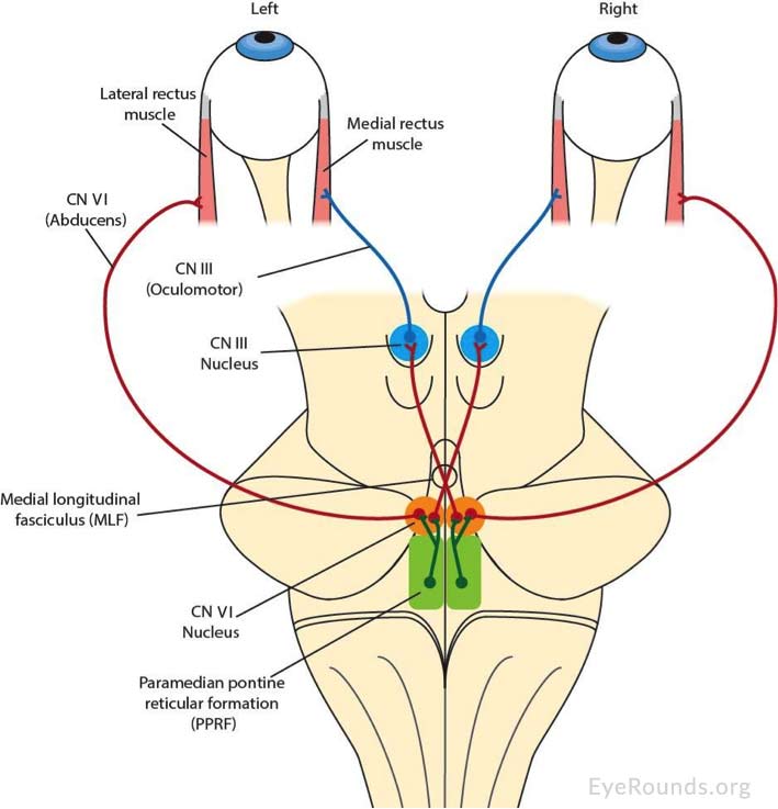
The medial longitudinal fasciculus (MLF) is a heavily myelinated tract connecting the abducens (CN VI) nucleus in the pons to the contralateral oculomotor (CN III) nucleus in the midbrain — the pathway that coordinates conjugate horizontal gaze, so that when one eye abducts, the other adducts in unison. A lesion in the MLF disconnects this coordination on one side.
The classic finding
- Impaired (slowed or absent) adduction of the eye ipsilateral to the lesion on attempted horizontal gaze toward the opposite side
- Dissociated (abducting) nystagmus of the contralateral eye as it abducts
- Convergence is preserved — this is the single most important distinguishing feature from a CN III palsy, since convergence uses a different pathway that bypasses the MLF. A CN III palsy impairs both adduction on lateral gaze and convergence; INO impairs only the lateral gaze component
- Can be unilateral or bilateral (bilateral INO is a strong red flag for MS, especially in a young patient)
Etiology by age — a genuinely useful pattern
| Population | Typical cause | Typical laterality |
|---|---|---|
| Young patients | Multiple sclerosis (demyelination) | Often bilateral |
| Older patients | Ischemic stroke (small vessel infarct) | Typically unilateral |
In one large review, infarction accounted for roughly 38% of cases (87% unilateral) and MS for roughly 34% (73% unilateral, meaning MS-related INO is more often bilateral than not-bilateral, but not exclusively so). Rarer causes include trauma, tumor, tentorial herniation, infection, and vasculitis.
A genuinely important current caveat: MRI can miss it
Because the MLF is extremely small and lacks contrast against surrounding brainstem tissue, conventional MRI is highly specific but poorly sensitive for INO — recent comparative work found MRI correctly identified only about 46% of INO cases confirmed by objective eye-movement recording (video-oculography), even though a positive MRI finding was highly reliable when present. In practical terms: a normal MRI does not rule out INO if the clinical exam is convincing — the diagnosis is fundamentally a clinical one, made at the bedside by careful observation of the adduction lag and dissociated nystagmus, not confirmed or excluded by imaging alone.
When lesions are visible, location can suggest cause: multiple MLF lesions favor demyelination (MS), while a lesion isolated to the mesencephalon favors ischemia.
Practical approach
- Confirm the finding carefully at the bedside — check both adduction lag and convergence specifically, since convergence preservation is what separates this from a CN III palsy
- In a young patient, especially with bilateral INO: this is a strong indication for MS workup (brain/spine MRI with contrast, consider CSF oligoclonal bands) even if the initial brainstem MRI doesn't show an obvious MLF lesion
- In an older patient with vascular risk factors and unilateral INO: evaluate as an ischemic event, with standard stroke workup
- Don't be reassured by a negative MRI in a clinically convincing case — treat the exam finding as pathognomonic and proceed with appropriate workup regardless
Key references: Internuclear Ophthalmoplegia, EyeWiki. | Kleinsorge MT, et al. MRI topography of lesions related to INO in MS or ischemic stroke. J Neuroimaging. 2021. | Lesion follows function: video-oculography compared with MRI to diagnose INO in MS. 2023.
📘 Want the exam-ready deep dive? This topic, and much more is covered in Ophthalmology Explorer (A High-Yield Clinical Reference for Ophthalmology Residents, Fellows & Board Exams) — available on Kindle. See all my books.
Want to know more about treatment? Read about Corneal Topography at Sadbhaav or book a consultation with Dr. Dhaval Patel.