Types of injections of the eyeball and types of corneal vascularization
Last updated: September 2026
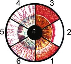
- superficial (conjunctival) injection
- deep (pericorneal) injection
- mixed injection
- superficial vascularization of the cornea
- deep vascularization of the cornea
- mixed corneal vascularization
Distinguishing the injection patterns
| Pattern | Appearance | Suggests |
|---|---|---|
| Superficial (conjunctival) | Bright red, most pronounced toward the fornices, moves freely with conjunctiva, blanches with topical phenylephrine | Conjunctivitis, superficial irritation, dry eye, allergic conjunctivitis |
| Deep (pericorneal/ciliary) | Violaceous/dusky red, concentrated around the limbus, does not move with conjunctiva, does not blanch with phenylephrine | Keratitis, iritis/anterior uveitis, acute angle closure, scleritis — treat as serious until proven otherwise |
| Mixed | Both patterns together | Often more severe or combined anterior segment disease |
Practical bedside tip: the phenylephrine blanching test is genuinely useful for rapid triage — superficial vessels blanch, deep ciliary vessels do not. A red eye that fails to blanch should be treated as a sign of potentially serious disease.
Corneal vascularization patterns
- Superficial: vessels continuous with conjunctival vessels, crossing the limbus superficially in a branching, tree-like pattern — seen in chronic contact lens wear, superficial keratitis, phlyctenular disease. Tends to regress once the cause is treated
- Deep: vessels from the anterior ciliary/episcleral circulation, running straight and parallel in the deep stroma — classic for interstitial keratitis (congenital syphilis, herpes). More persistent, and a recognized risk factor for graft rejection if keratoplasty is later needed
- Mixed: both patterns together, seen in more severe or longstanding corneal disease
📘 Want the exam-ready deep dive? This topic is covered in iNotes 2027: Basic Science & Optics (Ophthalmology Postgraduate Exam Notes) — available on Kindle. See all my books.
Experiencing symptoms like these? Learn about our Eye Department or book a consultation with Dr. Dhaval Patel.