Fundus Drawing: Colour Coding Chart
For: PG residents · Practising ophthalmologists
Key points in 60 seconds
- Drawn with the indirect ophthalmoscope, inverted, on a chart of circles for the equator and ora serrata.
- Red: attached retina, arteries, haemorrhages; breaks filled red and outlined in blue.
- Blue: detached retina, veins, ora serrata and lattice (blue cross-hatching).
- Green: vitreous opacities and haemorrhage; yellow: exudates and drusen.
- Black: pigment and laser or cryo scars; brown: choroidal detachment and naevi or melanoma.
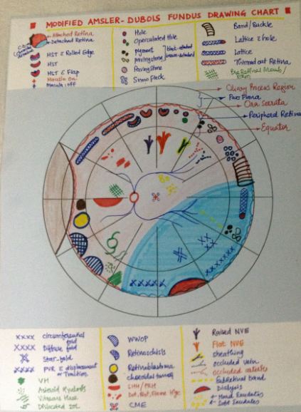
A fundus drawing records the retinal periphery, usually with an indirect ophthalmoscope, on a chart of concentric circles marking the equator and ora serrata. The drawing is made inverted, as seen through the condensing lens, and a standard colour code lets anyone read it at a glance.
Standard colour code
| Colour | What it shows |
|---|---|
| Red | Attached retina, retinal arteries, retinal haemorrhages, neovascularisation. Retinal breaks are filled red and outlined in blue. |
| Blue | Detached retina, retinal veins, outlines of breaks, the ora serrata, and lattice degeneration (blue cross-hatching outlined in blue) |
| Green | Vitreous and media opacities, vitreous haemorrhage, intraocular foreign bodies |
| Yellow | Exudates, drusen, retinal oedema |
| Black | Retinal pigment, pigment clumps, and pigmented laser or cryotherapy scars |
| Brown | Choroidal detachment, uveal tissue, choroidal naevi and melanoma |
Conventions vary slightly between units; follow your department's code. See also retinal detachment and Lincoff rules. For the anterior segment, see our corneal drawing colour chart.
Detailed fundus drawing chart
Companion chart: Corneal drawing: colour coding chart for slit-lamp drawings of the cornea.
How to cite this page
Patel D. Fundus Drawing: Colour Coding Chart. RPC Rounds (Sadbhaav Eye Clinic). Updated 2026. Available at: https://www.sadbhaavclinic.com/fundusdrawing.html. Accessed [date].
Experiencing symptoms like these? Learn about our Eye Department or book a consultation with Dr. Dhaval Patel.