Corneal Drawing: Colour Coding Chart
Last updated: November 2026
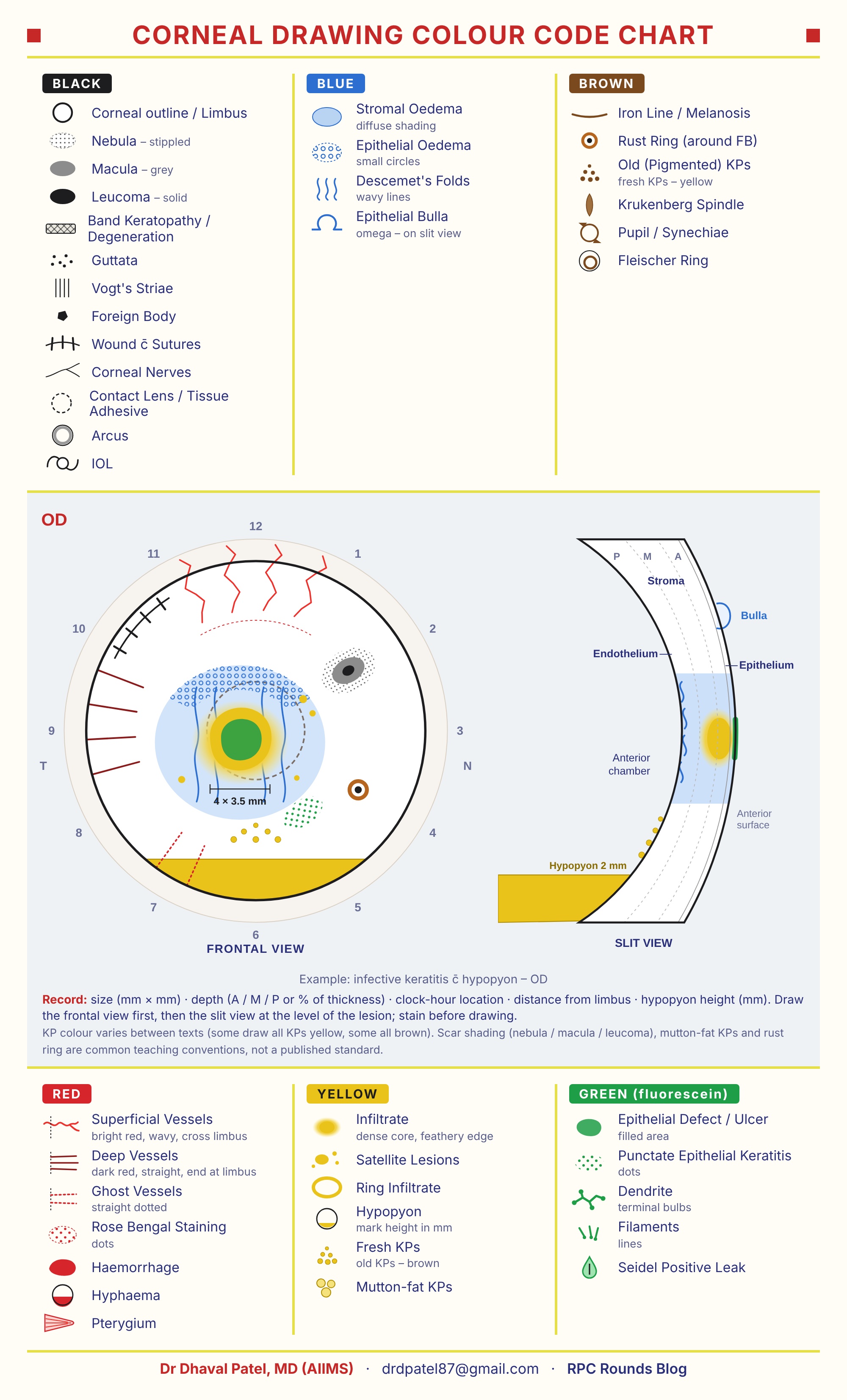
A corneal drawing records what you see at the slit lamp so that anyone can follow a keratitis, a scar or a graft from visit to visit. Draw two views: a frontal view of the cornea as a circle with clock hours, and a slit view (optical section) showing how deep the lesion goes. A shared colour code makes the drawing readable at a glance. This page is the corneal companion to our fundus drawing colour chart.
How to draw it
- Frontal view first. Draw the limbus as a circle and mark the clock hours, with temporal (T) and nasal (N) sides labelled for the eye you are drawing (OD or OS).
- Stain before you draw. Instil fluorescein and examine with the cobalt blue filter, so epithelial defects, punctate staining and a Seidel leak go in green at the same sitting.
- Draw the lesion to scale and write its size in millimetres (length × width), measured with the slit-beam length.
- Add the slit view at the level of the lesion: epithelium on the outside curve, endothelium on the inside, with the stroma divided into anterior, middle and posterior thirds (A / M / P).
- Record the anterior chamber: KPs, cells and the height of any hypopyon or hyphaema in millimetres.
Colour code
| Colour | What it shows |
|---|---|
| Black | Corneal outline and limbus; scars graded by density: nebula (stippled), macula (grey), leucoma (solid); band keratopathy; guttata; Vogt's striae; foreign body; wound with sutures; corneal nerves; contact lens or tissue adhesive (dashed outline); arcus; IOL |
| Blue | Oedema: stromal oedema as diffuse shading, epithelial oedema as small circles; Descemet's folds as wavy lines; epithelial bulla (an omega shape on the slit view) |
| Brown | Pigment: iron line and melanosis; rust ring around a foreign body; old (pigmented) KPs; Krukenberg spindle; pupil and synechiae; Fleischer ring |
| Red | Blood vessels and blood: superficial vessels bright red, wavy, crossing the limbus; deep vessels dark red, straight, ending at the limbus; ghost vessels as dotted lines; rose bengal staining as dots; haemorrhage; hyphaema; pterygium |
| Yellow | Inflammatory cells: infiltrate (dense core, feathery edge); satellite lesions; ring infiltrate; hypopyon (mark the height in mm); fresh KPs; mutton-fat KPs |
| Green | Fluorescein staining: epithelial defect or ulcer (filled area); punctate epithelial keratitis (dots); dendrite with terminal bulbs; filaments; Seidel-positive leak |
Telling look-alikes apart on the drawing
| Pair | How the drawing shows the difference |
|---|---|
| Superficial vs deep vessels | Superficial: bright red, wavy, branching, continuous with conjunctival vessels across the limbus. Deep: dark red, straight, parallel, stopping at the limbus. |
| Infiltrate vs scar | Infiltrate (yellow) has a dense core and a fuzzy, feathery edge and means active disease. A scar (black) has a sharp edge and graded density. |
| Epithelial defect vs infiltrate | Draw the green staining area and the yellow infiltrate separately and measure both: in healing, the defect closes before the infiltrate clears. |
| Fresh vs old KPs | Fresh KPs are yellow (active); old, pigmented KPs are brown. Mutton-fat KPs are large and greasy, typical of granulomatous uveitis. |
| Dendrite vs pseudodendrite | A herpetic dendrite has terminal bulbs and stains well (green). Pseudodendrites (healing abrasion, Acanthamoeba, zoster) lack true terminal bulbs. |
KP colour varies between textbooks: some draw all KPs yellow, others all brown. The scar shading (nebula, macula, leucoma), mutton-fat KPs and the rust ring symbols are common teaching conventions rather than a published standard. Follow your department's code and keep it consistent.
Detailed corneal drawing chart
The chart below shows every symbol, with a worked example of infective keratitis with hypopyon in the right eye (frontal and slit views).
Related: Fundus drawing colour chart, Microbial keratitis, Dendrites, Cornea hub, All downloads.
Experiencing symptoms like these? Learn about our Eye Department or book a consultation with Dr. Dhaval Patel.