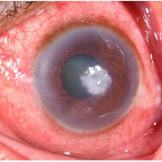
Microbial Keratitis: A Workup Approach
Last updated: September 2026
Microbial keratitis is a true ophthalmic emergency where the diagnostic workup and empirical treatment happen in parallel, not sequentially — waiting for culture results before starting treatment risks irreversible corneal scarring and vision loss.
Epidemiology: bacteria dominate, but know the outliers
Recent large series consistently show bacteria as the leading cause (roughly 52-90% of culture-positive cases depending on region and population), followed by fungi and Acanthamoeba. Pseudomonas aeruginosa and Staphylococcus species are frequently implicated bacterial isolates; Fusarium is the leading fungal cause in many warm, humid, agricultural regions. Ocular trauma increases microbial keratitis risk roughly 5-fold, and agricultural activity roughly 1.3-fold — both worth asking about specifically in history-taking.
The workup: corneal scraping remains central
- Do not delay antibiotics to "clear" the culture first — the older practice of withholding antibiotics for 12 hours before scraping to improve culture yield is no longer recommended by most practitioners; obtain the scraping, then start empirical treatment immediately
- Smears: Gram stain (bacteria), Giemsa, acid-fast stain (mycobacteria/Nocardia), and KOH with calcofluor white (fungi and Acanthamoeba) — KOH/calcofluor white has notably higher sensitivity for fungi (~90%) and Acanthamoeba (~84%) than Gram stain has for bacteria (~57%), a genuinely useful fact when interpreting a negative smear
- For mild, small peripheral infiltrates (<1-2mm), many practitioners reasonably defer scraping and treat empirically, particularly where lab resources are limited
- Confocal microscopy is an increasingly valuable adjunct, allowing direct in-vivo visualization of fungal filaments and Acanthamoeba cysts in the stroma without waiting for culture
- Impression cytology (a cellulose acetate filter applied to the cornea rather than a blade) is an emerging alternative sample-collection method, with comparable or superior isolation rates to traditional scraping in recent large comparative studies
Clinical clues that should raise suspicion for Acanthamoeba
Acanthamoeba is rarely culture-positive on routine scraping, so recognizing its distinct clinical pattern matters more than lab confirmation early on:
- Pseudodendrites (can be mistaken for HSV)
- Perineural infiltrates (radial keratoneuritis) — a relatively specific finding
- Ring-shaped infiltrate, particularly if it develops later in a case initially presumed bacterial or fungal
- Contact lens wear, especially with tap water exposure or poor lens hygiene
- Disproportionate pain relative to clinical signs early in the disease
- Longer symptom duration before presentation, and prior topical antibiotic use without improvement
- A non-healing, worsening ulcer despite appropriate antibacterial/antiviral therapy should always prompt reconsidering Acanthamoeba rather than simply escalating the presumed-bacterial or presumed-viral treatment
Empirical treatment paradigm
- Presumed bacterial: broad-spectrum fortified antibiotics covering both Gram-positive and Gram-negative organisms — fortified cefazolin plus a fluoroquinolone (e.g., moxifloxacin) is a commonly used combination
- Presumed fungal: topical natamycin 5% (particularly effective for filamentous fungi like Fusarium) or voriconazole; oral voriconazole may be added for deeper/severe disease
- Presumed Acanthamoeba: topical PHMB (polyhexamethylene biguanide) and/or propamidine, often combined with a fluoroquinolone for bacterial co-coverage; prolonged treatment (often continued 2+ weeks beyond symptom resolution) is standard given the cyst stage's resistance to treatment
- Therapy is refined once culture and sensitivity results return, narrowing from broad empirical coverage to targeted treatment
- Fungal keratitis carries the worst prognosis of the three in comparative outcome data — a meaningfully higher proportion require surgical intervention (roughly 51% vs. 43% for bacterial) and achieve a healed scar less often, reinforcing why accurate early fungal diagnosis matters so much
Key references: Five-Year Analysis of Microbial Keratitis Incidence and Sensitivity, South West England. 2025. | Review of epidemiological features, diagnosis and treatment outcome of microbial keratitis. | Emerging diagnostic modalities in microbial keratitis: beyond culture and smear. 2025.
📘 Want the exam-ready deep dive? This topic is covered in iNotes 2027: Cornea and Refractive (Ophthalmology Postgraduate Exam Notes) — available on Kindle. See all my books.
Want to know more about treatment? Read about Corneal Topography at Sadbhaav or book a consultation with Dr. Dhaval Patel.