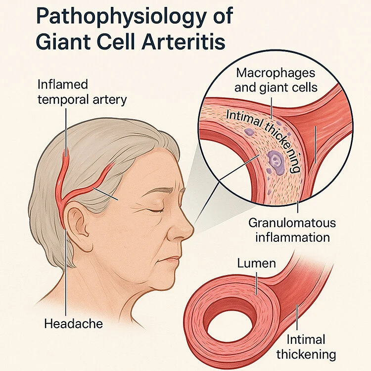
Giant Cell Arteritis: The Emergency You Can't Miss
Last updated: September 2026
Giant cell arteritis (GCA) is the ophthalmic emergency you cannot afford to miss in any patient over 50 with sudden vision loss — untreated, it risks bilateral, permanent blindness within days, yet it responds dramatically to prompt high-dose steroids started even before biopsy confirmation.
Why the urgency
- GCA causes ischemic damage to the ophthalmic and posterior ciliary arteries, most commonly presenting as arteritic anterior ischemic optic neuropathy (AAION)
- Fellow-eye involvement can occur within days if untreated — this is precisely why steroids are started empirically on clinical suspicion, without waiting for biopsy results
- Vision loss from GCA is typically irreversible once it occurs — treatment is about preventing further loss, not reversing damage already done
Classic symptoms to actively ask about
- New temporal headache
- Jaw or tongue claudication (pain with chewing that improves with rest — highly specific)
- Scalp tenderness (classically, pain when combing hair)
- Sudden, painless monocular vision loss
- Systemic features of polymyalgia rheumatica: shoulder and hip girdle stiffness, worse in the morning
- Amaurosis fugax (transient vision loss) can precede permanent loss — a warning sign not to dismiss
Current diagnostic framework: 2022 ACR/EULAR classification criteria
This replaced the older 1990 ACR criteria, which performed poorly for the large-vessel GCA subtype (sensitivity as low as 37% in that group). The new criteria weight both clinical features and modern imaging:
| Criterion | Points |
|---|---|
| Age ≥50 years at diagnosis | Mandatory absolute requirement |
| Positive temporal artery biopsy, or halo sign on temporal artery ultrasound | +5 |
| ESR ≥50mm/hr or CRP ≥10mg/L | +3 |
| Sudden visual loss | +3 |
| Morning stiffness in shoulders/neck, jaw or tongue claudication, new temporal headache, scalp tenderness, abnormal temporal artery on exam, bilateral axillary involvement on imaging, FDG-PET activity throughout the aorta | +2 each |
A cumulative score ≥6 classifies GCA, with strong performance in validation (AUC 0.91, sensitivity 87%, specificity 95%).
A genuinely current shift: ultrasound as a real alternative to biopsy
Temporal artery biopsy has long been the diagnostic reference standard — but recent evidence increasingly supports temporal artery ultrasound (looking for the "halo sign") as a real alternative, weighted equally to biopsy in the 2022 criteria. EULAR now recommends ultrasound as a first-line diagnostic tool for suspected large-vessel vasculitis, avoiding an invasive procedure in many patients when ultrasound is positive and clinical suspicion is high. That said, ACR/Vasculitis Foundation guidance still favors biopsy as the reference standard where ultrasound expertise isn't readily available — practice varies by center and access to trained sonographers.
Management principle
- Start high-dose systemic corticosteroids immediately on clinical suspicion — do not wait for biopsy or ultrasound results if the clinical picture is concerning, especially with any visual symptoms
- Biopsy (or ultrasound) can still be diagnostic even after several days of steroid treatment, so starting treatment doesn't meaningfully compromise the ability to confirm the diagnosis
- Long-term management requires rheumatology co-management given the risk of relapse and steroid-related morbidity with prolonged treatment courses
Key references: 2022 ACR/EULAR classification criteria for giant cell arteritis. Ann Rheum Dis. 2022. | Giant cell arteritis: reviewing the advancing diagnostics and management. Eye. 2023.
📘 Want the exam-ready deep dive? This topic, and much more is covered in Ophthalmology Explorer (A High-Yield Clinical Reference for Ophthalmology Residents, Fellows & Board Exams) — available on Kindle. See all my books.
Want to know more about treatment? Read about Corneal Topography at Sadbhaav or book a consultation with Dr. Dhaval Patel.