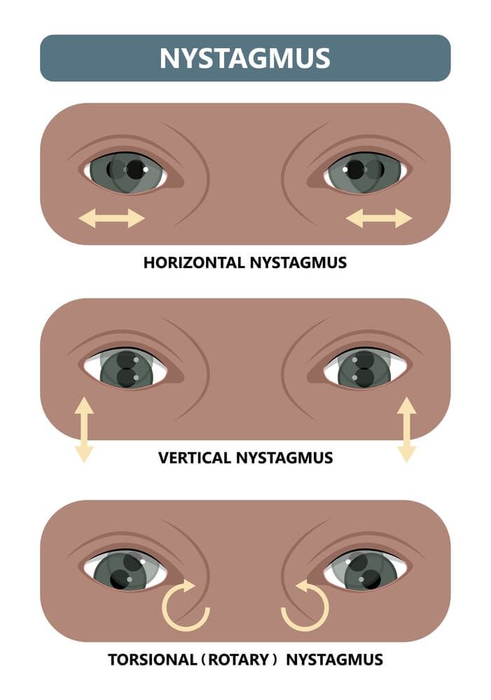
Nystagmus: A Classification Approach
Last updated: September 2026
Nystagmus is a description, not a diagnosis — rhythmic, involuntary oscillation of the eyes. The real clinical work is characterizing the waveform, timing of onset, and specific pattern, since each of these narrows the differential dramatically.
Step 1: Physiologic or pathologic?
Brief, low-amplitude forms — end-point nystagmus (at extremes of gaze) and optokinetic nystagmus (in response to moving stripes) — are normal findings in healthy people and resolve when the stimulus is removed. Persistent oscillation lasting more than a few seconds, or any oscillation outside these specific normal contexts, should be considered pathologic until proven otherwise.
Step 2: Age at onset — infantile vs. acquired
| Infantile (congenital) | Acquired | |
|---|---|---|
| Onset | Birth to 6 months | After 6 months, any age |
| Oscillopsia (perceived visual shaking) | Absent — the brain has adapted since birth | Often present — genuinely disturbing to the patient, and its presence is itself a useful clue that the nystagmus is acquired |
| Typical pattern | Bilateral, conjugate, horizontal; pendular or jerk with an accelerating slow phase; often dampens with convergence and shows a null point (gaze position of minimum intensity) | Variable by cause — direction and waveform carry strong localizing value |
Infantile nystagmus syndrome (INS) can be idiopathic or associated with sensory deficits — albinism, congenital cataract, retinal dystrophies, or optic nerve hypoplasia — so any infant with nystagmus needs a full ocular exam looking for an underlying sensory cause, not just observation.
Special patterns with strong localizing value
The AAO explicitly flags these forms as requiring imaging and specialist workup whenever seen — they are rarely benign:
- Downbeat nystagmus — craniocervical junction pathology (Arnold-Chiari malformation), cerebellar disease, or drug toxicity (lithium, anticonvulsants)
- Upbeat nystagmus — brainstem or cerebellar lesions
- Seesaw nystagmus — one eye rises and intorts while the other falls and extorts; classically parasellar/chiasmal lesions
- Periodic alternating nystagmus — direction reverses every 1-2 minutes; craniocervical junction or cerebellar disease
- Convergence-retraction nystagmus — part of dorsal midbrain (Parinaud) syndrome, elicited on attempted upgaze
- Acquired pendular nystagmus — regular, sinusoidal oscillation; multiple sclerosis is the most common cause, also seen with brainstem lesions, Whipple's disease, or drug toxicity
- Oculomasticatory myorhythmia — rhythmic convergence-divergence movements paired with jaw muscle contraction — pathognomonic specifically for Whipple's disease
Latent nystagmus — a distinct pediatric pattern worth knowing
A conjugate jerk nystagmus, fast phase toward the fixing eye, classically seen with congenital esotropia or after infantile esotropia surgery — thought to reflect abnormal binocular development. Amplitude increases when one eye is covered ("manifest latent nystagmus" when it becomes constantly visible). Clinically relevant for accurate visual acuity testing in these children: use a technique that doesn't fully occlude the fellow eye (e.g., a high-plus blur lens or polarized vectograph) to avoid artificially worsening the nystagmus and understating true acuity.
Treatment principle
Treatment targets the underlying cause where one exists (e.g., MS-directed therapy for demyelinating disease). For acquired pendular nystagmus specifically, pharmacologic options with the most robust trial evidence are gabapentin and memantine — both have undergone more rigorous treatment trials than other proposed agents, based on the pathophysiologic rationale of the oscillation.
Key references: Nystagmus in Clinical Practice: From Diagnosis to Treatment. Clin Ophthalmol. 2025. | Nystagmus Types, StatPearls (2025). | AAO Clinical Guidelines: Childhood Nystagmus Workup.
📘 Want the exam-ready deep dive? This topic, and much more is covered in Ophthalmology Explorer (A High-Yield Clinical Reference for Ophthalmology Residents, Fellows & Board Exams) — available on Kindle. See all my books.
Want to know more about treatment? Read about Corneal Topography at Sadbhaav or book a consultation with Dr. Dhaval Patel.