
Angle Closure Glaucoma Definitions
Last updated: October 2026
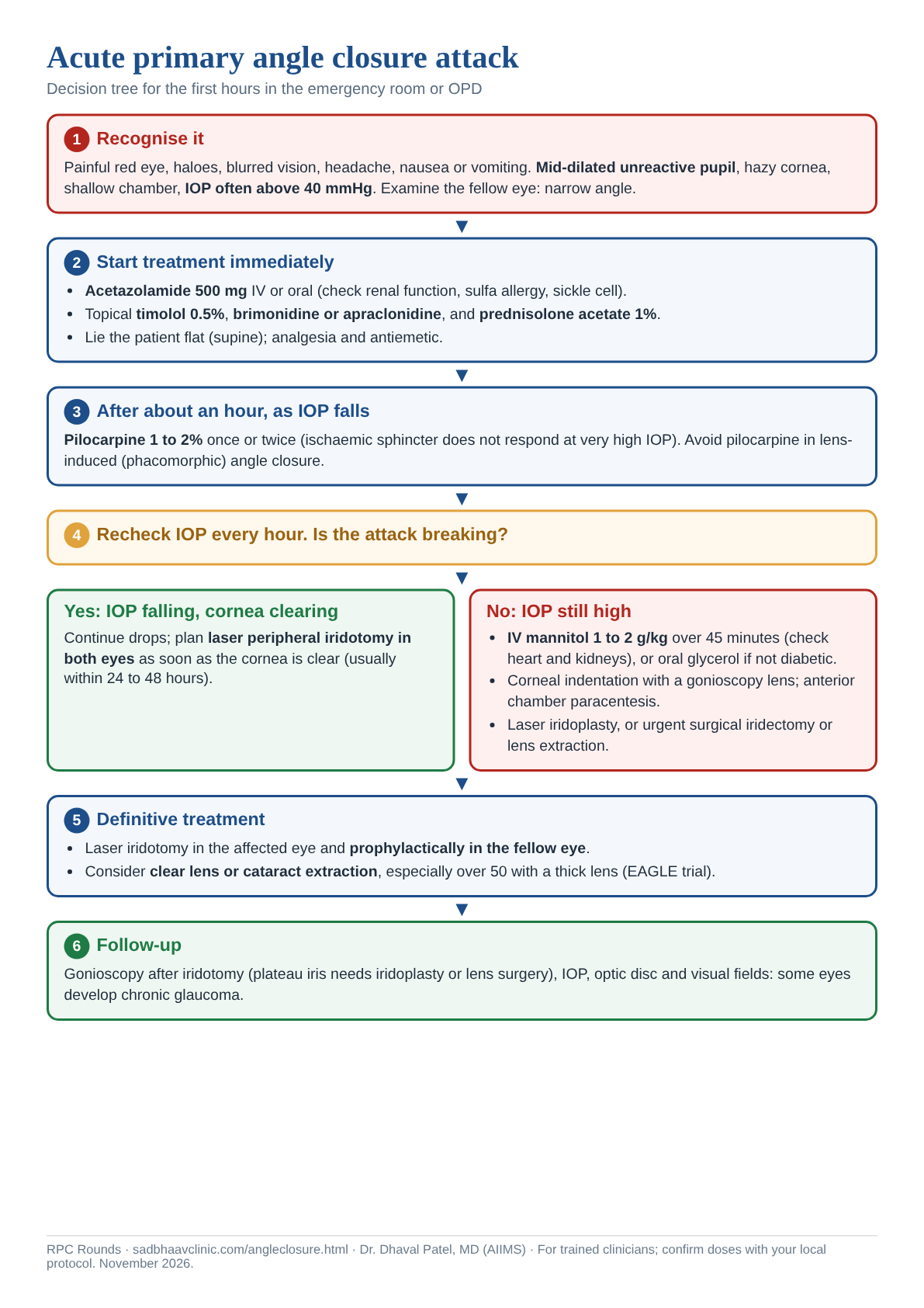
Decision tree: Acute angle closure attack
A one-page, printable step-by-step flowchart for the emergency room or OPD.
The ISGEO (International Society for Geographical and Epidemiological Ophthalmology) classification remains the standard framework for staging angle closure disease — precise terminology here matters because it directly determines management.
Definitions
| Term | Gonioscopy | Other findings |
|---|---|---|
| Primary angle-closure suspect (PACS) | Posterior trabecular meshwork iridotrabecular contact (ITC) in three or more quadrants, but no peripheral anterior synechiae (PAS) | Normal IOP, optic disc, and visual field. A lower threshold — even two quadrants of ITC, or a narrow angle approach of ~20° — may still justify the diagnosis in some patients showing evidence of intermittent angle closure |
| Primary angle-closure (PAC) | Three or more quadrants of ITC, with raised IOP and/or PAS, or excessive pigment smudging on the trabecular meshwork | Normal optic disc and visual field — the key distinction from PACG is the absence of glaucomatous optic neuropathy |
| Primary angle-closure glaucoma (PACG) | ITC in three or more quadrants | Glaucomatous optic neuropathy present — this is what separates PACG from PAC |
Current diagnostic addition: AS-OCT alongside gonioscopy
While gonioscopy remains the clinical reference standard for angle assessment, anterior segment OCT is now widely used as a complementary, non-contact tool — particularly useful for objective, reproducible angle measurements and for patients where gonioscopy is difficult (photophobia, corneal haze, or poor cooperation).
Important current shift in first-line management
Randomized trial evidence (including the EAGLE trial) has shown that in select patients — people aged 50 or older with primary angle closure and IOP of 30 mmHg or more, or with PACG (acute attacks were excluded) — clear lens extraction can outperform laser peripheral iridotomy (LPI) as an initial treatment, by permanently deepening the anterior chamber rather than simply creating a pressure-equalizing bypass. This has meaningfully shifted first-line thinking in some angle-closure scenarios, especially when a visually significant cataract already coexists — worth knowing as current evidence-based practice rather than defaulting reflexively to "LPI for everyone."
2026 update: what ZAP and ANA-LIG changed
| Trial | Finding | Practice point |
|---|---|---|
| ZAP (2019, China) | Prophylactic iridotomy in one eye of PACS patients: progression to PAC was low (about 4 vs 8 per 1000 eye-years, under 1% a year), so the absolute benefit was small | Laser iridotomy is not routine for PACS; consider it for higher risk (narrow angles needing dilation, poor access to care, symptoms) |
| ANA-LIG (Singapore) | Similar low progression rates after iridotomy in PACS | Supports a selective approach |
| EAGLE (2016) | Clear lens extraction beat iridotomy for PAC with IOP ≥30 mmHg or PACG | Consider lensectomy early in PAC/PACG |
| Terminology | PACS is now iridotrabecular contact of 180° or more (was 270°) | See ISGEO in the classifications page |
Sources for this update: He M, et al. Laser peripheral iridotomy for the prevention of angle closure (ZAP). Lancet. 2019;393:1609-1618 | Azuara-Blanco A, et al. Clear-lens extraction versus laser iridotomy (EAGLE). Lancet. 2016;388:1389-1397.
Revise with flip cards
Key references: Weinreb RN, et al. Primary angle-closure disease: ISGEO classification. | Day AC, et al. EAGLE trial: clear lens extraction for primary angle closure glaucoma. Lancet.
📘 Want the exam-ready deep dive? This topic is covered in iNotes 2027: Glaucoma (Ophthalmology Postgraduate Exam Notes) — available on Kindle. See all my books.
Want to know more about treatment? Read about Glaucoma Treatment at Sadbhaav or book a consultation with Dr. Dhaval Patel.