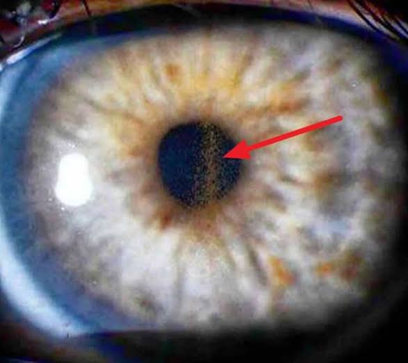
Pigment Dispersion Syndrome: A Mechanical Story
Last updated: September 2026
Pigment dispersion syndrome (PDS) has a genuinely elegant, mechanical explanation that directly points to its treatment — understanding the "reverse pupillary block" mechanism makes the whole condition click into place rather than being a list of memorized signs.
The mechanism: reverse pupillary block
In susceptible eyes (typically young, myopic, with a deep anterior chamber), the mid-peripheral iris bows backward into a concave configuration, bringing it into contact with the anterior lens zonules. This concavity is thought to result from a pressure differential — aqueous pressure anterior to the iris exceeds that posterior to it, the reverse of the usual relationship — triggered or worsened by blinking (which forces small aqueous aliquots forward) and accommodation. The mechanical rubbing between the posterior iris pigment epithelium and the zonules liberates pigment granules, which then disperse throughout the anterior segment.
The classic clinical triad
- Krukenberg spindle — a vertical, fusiform band of pigment on the corneal endothelium (from convection currents depositing pigment centrally on the posterior cornea). Note: this alone is not pathognomonic — it can also occur in pseudoexfoliation, uveitis, and after trauma
- Radial, mid-peripheral iris transillumination defects — spoke-like defects best seen with deliberate retroillumination technique, corresponding to the areas of iridozonular rubbing
- Dense, homogeneous trabecular meshwork pigmentation on gonioscopy — the homogeneous distribution pattern helps distinguish PDS from pseudoexfoliation, which tends to show patchier pigmentation
Other findings: Zentmayer's ring (pigment on the posterior lens surface) and Scheie's stripe.
PDS vs. pigmentary glaucoma: the distinction that matters
PDS describes the clinical triad above without optic nerve damage or visual field loss, even if IOP happens to be elevated. Pigmentary glaucoma is diagnosed once glaucomatous cupping and/or field loss develops. Roughly 10% of PDS patients progress to pigmentary glaucoma within 5 years, rising to a broader estimate of 25-50% over the longer term — meaning most, but not all, PDS eventually needs active glaucoma management, and ongoing monitoring is essential even in initially "benign" PDS.
A practical clinical pearl: exercise-related IOP spikes
Vigorous exercise (particularly activities involving pupil dilation/accommodation, like running) can provoke acute pigment liberation and transient IOP spikes in PDS patients — worth specifically asking about exercise-related visual symptoms (halos, blurring) and considering post-exercise IOP measurement if this history is present.
Current management approach
- PDS without glaucoma: monitoring is the mainstay — regular IOP checks, optic nerve OCT, and visual field testing to catch conversion to pigmentary glaucoma early
- Laser peripheral iridotomy (LPI): directly targets the reverse pupillary block mechanism — by equalizing pressure between the anterior and posterior chambers, LPI flattens the concave iris configuration, physically separating it from the zonules and halting further pigment release. This is a genuinely mechanism-based treatment, not just symptomatic IOP control, when reverse pupillary block is confirmed (e.g., on ultrasound biomicroscopy or AS-OCT)
- Once pigmentary glaucoma develops: managed similarly to other open-angle glaucomas — topical IOP-lowering medication, laser trabeculoplasty (effective, though some studies suggest results may be less durable in younger PDS patients due to higher pigment turnover), and incisional glaucoma surgery reserved for refractory cases
- Argon laser iridoplasty is an emerging, less commonly used adjunct specifically studied for altering iris configuration in PDS
Key references: Pigmentary Glaucoma and Pigment Dispersion Syndrome, EyeWiki (2026). | Pigment Dispersion Syndrome and Pigmentary Glaucoma: New Clinical Gradation and Current Therapeutic Strategies. 2025. | Pigment dispersion syndrome and pigmentary glaucoma: the basics. EyeWorld. 2025.
📘 Want the exam-ready deep dive? This topic is covered in iNotes 2027: Glaucoma (Ophthalmology Postgraduate Exam Notes) — available on Kindle. See all my books.
Want to know more about treatment? Read about Corneal Topography at Sadbhaav or book a consultation with Dr. Dhaval Patel.