Retinopathy of Prematurity: ICROP3 Update
Last updated: September 2026
Retinopathy of prematurity (ROP) screening exists because timely treatment genuinely prevents blindness — but the classification system used to describe what you're seeing was substantially revised in 2021, and residents trained on older teaching should know what changed.
The three core descriptors (unchanged since 1984)
- Zone — location of disease relative to the optic disc (Zone I = posterior pole, most severe; Zone II = mid-periphery; Zone III = far periphery, least severe)
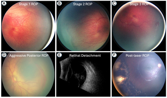
- Stage — appearance at the vascular-avascular junction (Stage 1 = demarcation line; Stage 2 = ridge; Stage 3 = ridge with extraretinal fibrovascular proliferation; Stage 4 = partial retinal detachment; Stage 5 = total retinal detachment)
- Circumferential extent — measured in clock hours
What ICROP3 (2021) actually changed
The third edition was needed because of subjectivity concerns in earlier criteria, newer imaging technology, and — most importantly — anti-VEGF therapy producing regression/reactivation patterns the older system wasn't built to describe:
- "Posterior Zone II" is now formally recognized — a region just outside Zone I that behaves more aggressively than the rest of Zone II, closer in risk to Zone I disease
- "Notch" — describes focal posterior extension of disease into a more posterior zone than the rest of the circumference, which can be easy to miss if you're only recording the single worst zone overall
- Stage 5 is now subcategorized into 5A/5B/5C based on the configuration of the retinal detachment — relevant for surgical planning and prognosis
- "Aggressive ROP" replaces "aggressive posterior ROP" — reflecting recognition that this aggressive phenotype isn't confined to the posterior pole and can occur in larger, more mature preterm infants, especially in resource-limited settings where the disease pattern doesn't always fit the classic textbook picture
- Plus disease is now recognized as existing on a continuous spectrum rather than a strict present/absent binary, with "pre-plus" as an intermediate category
- Detailed new guidance on regression and reactivation patterns after anti-VEGF treatment — since these eyes can reactivate later in a way ablative laser-treated eyes typically don't, requiring a different, often longer follow-up schedule
- Long-term sequelae are now explicitly described: late retinal detachment, retinoschisis, persistent avascular retina, macular anomalies, and secondary angle-closure glaucoma — reinforcing that ROP follow-up doesn't simply end once acute disease regresses
Screening principles
- Screening criteria are based on gestational age and birth weight, refined by evidence like the postnatal growth and ROP (G-ROP) validation studies that help identify which infants can safely be screened less intensively
- Telemedicine using wide-angle retinal imaging is increasingly used, particularly to extend screening access in low-resource settings and reduce the burden of in-person binocular indirect ophthalmoscopy for every at-risk infant
- AI-assisted ROP screening is an active area of development, though still early-stage and not yet a replacement for expert examination
Why this matters for cross-linking your other differentials
ROP is one of the classic causes of both leukocoria (from stage 4-5 detachment) and needs to be distinguished from other causes of white pupil in a premature infant, particularly persistent fetal vasculature and congenital cataract.
Key references: International Classification of Retinopathy of Prematurity, Third Edition. Ophthalmology. 2021;128:e51-e68. | Screening and surveillance for ROP: a Wilson and Jungner framework approach. 2023.
📘 Want the exam-ready deep dive? This topic, and much more is covered in Ophthalmology Explorer (A High-Yield Clinical Reference for Ophthalmology Residents, Fellows & Board Exams) — available on Kindle. See all my books.
Want to know more about treatment? Read about Corneal Topography at Sadbhaav or book a consultation with Dr. Dhaval Patel.