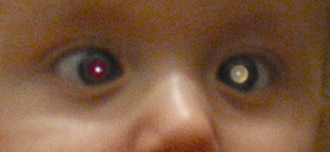
Leukocoria: A Differential
Last updated: September 2026
Leukocoria — a white pupillary reflex instead of the normal red reflex — is a sign, not a diagnosis, but it's the sign no ophthalmologist can afford to be casual about, since retinoblastoma sits at the top of every differential list until excluded.
Why the urgency
Retinoblastoma is diagnosed in roughly 47% of children referred with leukocoria to tertiary centers, and leukocoria is the presenting sign in the majority (32-73%) of retinoblastoma cases — but it's genuinely a sign shared by many other conditions, several of which are also vision- or life-threatening in their own right.
The core differential
| Cause | Distinguishing clue |
|---|---|
| Retinoblastoma | Calcification on ultrasound (seen in ~90% of cases) — highly specific; elevated mass on fundus exam; may have associated strabismus |
| Congenital cataract | Opacity visible in the lens itself on slit lamp exam, anterior to the retina — no mass behind it |
| Persistent fetal vasculature (PFV/PHPV) | Usually unilateral, microphthalmic eye, retrolental fibrovascular stalk visible, often with an elongated ciliary process ("dragging") |
| Coats disease | Telangiectatic retinal vessels with subretinal lipid exudation, typically unilateral, boys affected more often, no calcification on ultrasound (a key point distinguishing it from retinoblastoma) |
| Retinopathy of prematurity (stage 4-5) | History of prematurity is the key clue — see our ROP page |
| Retinal detachment (other causes) | Thorough fundus exam for a mass lesion or seeds to differentiate from retinoblastoma-associated detachment |
| Toxocariasis / other posterior inflammation | History of pica or geophagia, exposure to puppies; inflammatory signs, granuloma |
| Uveal coloboma | Sharply demarcated, glistening white, bowl-shaped excavation — no elevated mass, distinguishing it from tumor |
| Familial exudative vitreoretinopathy (FEVR) | Peripheral avascular retina, often bilateral, family history — genetically distinct from ROP but can look similar |
| Simple asymmetric refractive error / strabismus | Can produce an asymmetric red reflex (Bruckner test) without true leukocoria — always worth ruling out as a benign mimic before extensive workup |
Practical workup sequence
- Ocular ultrasound (B-scan) in every case — inexpensive, non-invasive, and highly specific for detecting the calcifications characteristic of retinoblastoma
- Dilated fundus exam under anesthesia if needed, given the exam demands in an infant
- MRI (avoid CT where possible, to limit radiation exposure in a child with a possible heritable cancer predisposition) to assess for optic nerve or intracranial extension if retinoblastoma is confirmed or strongly suspected — see our Retinoblastoma Genetics page for what happens next if confirmed
- Biopsy is not performed for suspected retinoblastoma — the risk of extraocular tumor seeding makes this contraindicated; diagnosis relies entirely on clinical exam and imaging
A practical real-world detection tip
A substantial proportion of retinoblastoma cases (up to 80% in one study) are first noticed by parents in recreational flash photographs, where the tumor produces a white reflex instead of the normal "red-eye" — worth mentioning to families as something to watch for, since this often prompts presentation earlier than a routine well-child exam might catch it.
Key references: A Stepwise Approach to Leukocoria, AAO EyeNet. | Educational Case: Retinoblastoma. 2025. | Moran CORE: Leukocoria in Children.
📘 Want the exam-ready deep dive? This topic, and much more is covered in Ophthalmology Explorer (A High-Yield Clinical Reference for Ophthalmology Residents, Fellows & Board Exams) — available on Kindle. See all my books.
Want to know more about treatment? Read about Corneal Topography at Sadbhaav or book a consultation with Dr. Dhaval Patel.