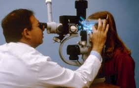
Slit Lamp Laser Lens
Last updated: September 2026
Choosing the right contact lens for slit-lamp laser delivery affects magnification, field of view, and image orientation — all of which directly influence spot sizing calculations and treatment accuracy. Knowing these differences cold is essential before ever sitting at the laser.
Goldmann three-mirror lens
- The classic, most widely taught panretinal/PRP lens
- Central lens for posterior pole; three angled mirrors (equatorial, mid-peripheral, and peripheral/gonioscopy mirror) to access progressively more peripheral retina and the angle
- Requires a coupling agent (methylcellulose) and direct corneal contact
- Image is inverted and reversed through the mirrors — orientation must be mentally corrected during treatment
Wide-field panfundoscopic lenses (e.g., Rodenstock, Mainster wide-field)
- Provide a much larger field of view than the Goldmann lens — allow more retina to be treated per laser application, speeding up PRP sessions
- Higher lens magnification typically means the actual retinal spot size is larger than the machine's dial-in setting — this is critical to account for, since under-correcting spot size leads to unintentionally intense, higher-power-density burns
- Preferred by many retina specialists specifically for PRP given the time efficiency
Macular/focal treatment lenses (e.g., Mainster focal/grid, Area Centralis)
- Higher magnification, smaller field of view — designed for precision work at the posterior pole (focal/grid laser for diabetic macular edema, focal treatment near vascular lesions)
- The higher magnification means the actual retinal spot size is smaller than the dial-in setting — the opposite correction direction from wide-field lenses
Key practical principle: lens magnification changes real spot size
Every contact lens has its own magnification factor relative to the laser's dialed-in spot size — this is not optional knowledge, since using the wrong correction factor directly changes treatment intensity at the retina:
- Wide-field/panfundoscopic lenses: real retinal spot size is larger than the dial-in setting
- High-magnification macular lenses: real retinal spot size is smaller than the dial-in setting
- Always confirm the specific magnification factor for the exact lens model in use — these vary by manufacturer and are provided in the lens documentation
Practical tips
- Always use adequate coupling gel to avoid air bubbles that distort the view and can scatter laser energy unpredictably
- Confirm image orientation (especially with three-mirror lenses) before starting treatment near critical structures like the fovea or major vessels
- Recheck spot size correction whenever switching lenses mid-session — a common, avoidable source of inconsistent burn intensity
📘 Want the exam-ready deep dive? This topic, and much more is covered in Ophthalmology Explorer (A High-Yield Clinical Reference for Ophthalmology Residents, Fellows & Board Exams) — available on Kindle. See all my books.
Want to know more about treatment? Read about Retina Treatment at Sadbhaav or book a consultation with Dr. Dhaval Patel.