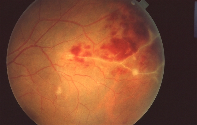
Eales Disease
Last updated: September 2026
Important Percentages
- NVI: 0.5-0.8 %
- NVD: 10-18 %
- NVE: 50 %
- Sec RRD: 8-11 %
Proliferative vascular retinopathy mimicking eales
- SYSTEMIC: DM, Sarcoidosis, Sickle
- OCULAR: BRVO, CRVO, Coats, Pars Planitis, ROP
Main causes of vision loss in Eales’ Disease
- SUDDEN
- Vitreous hemorrhage (NVE, Inflammatory BRVO, NVD)
- Secondary rhegmatogenous retinal detachment
GRADUAL
- Macular edema
- Traction retinal detachment involving posterior pole
- Disc edema
- Multiple vitreous membranes
- Cataract
- Glaucoma – NVG/Ghost cell
- Optic atrophy
- Epimacular membrane
Main indications for vitrectomy in Eales’ disease
- Non-resolving vitreous hemorrhage
- Traction retinal detachment involving posterior pole
- Multiple vitreous membranes
- Combined traction and rhegmatogenous retinal detachment
- Vitreous hemorrhage associated with hemolytic or ghost-cell glaucoma
- Macular pucker / Epimacular membrane
- Vitreous hemorrhage associated with rubeosis
Management
- If B/L: Systemic Steroids
- If U/L:
- <2 quadrent: PST
- >2 quadrent: Systemic Steroids
Current understanding of pathogenesis
Eales' disease remains a diagnosis of exclusion, but the leading current hypothesis implicates hypersensitivity to Mycobacterium tuberculosis antigens — supported by molecular evidence of innate immune activation (CD16+ monocytes with upregulated TLR-2) driving a pro-inflammatory cascade (elevated TNF-α and IL-6) that promotes endothelial activation and MMP-9-mediated extracellular matrix breakdown. A significantly elevated vitreous VEGF/PEDF ratio shifts the retinal environment decisively toward neovascularization — the same downstream pathway shared with other ischemic retinopathies, which is why anti-VEGF has a role here despite the distinct underlying trigger.
Current stage-based treatment approach
| Stage | Current approach |
|---|---|
| Inflammatory (periphlebitis) | Systemic corticosteroids, combined with antitubercular therapy (ATT) when a rigorous TB workup supports it |
| Ischemic (capillary non-perfusion) | Panretinal photocoagulation of ischemic areas to pre-empt neovascularization — timely laser before neovascularization develops improves prognosis |
| Proliferative (neovascularization) | Laser photocoagulation remains central; intravitreal anti-VEGF can be added to accelerate regression of active neovascularization; vitrectomy for non-resolving vitreous hemorrhage or tractional detachment |
Important current caveats
- A positive IGRA or Mantoux test alone does not mandate ATT — the decision must incorporate HRCT chest findings, the angiographic phenotype, and clinical course, calibrated to regional TB epidemiology. Reflexively starting ATT on a positive TB test alone is not the current standard of care
- Anti-VEGF requires caution in eyes with established fibrovascular proliferation — while effective at regressing active neovascularization, anti-VEGF can accelerate fibrosis and traction in eyes with mature fibrovascular membranes, so timing and case selection matter
- Laser should not be applied over areas of active vasculitis — treating inflamed, not just ischemic, retina is a recognized pitfall
- Newer anti-VEGF/anti-angiopoietin-2 agents (e.g., faricimab) have been used successfully as an adjunct to vitrectomy in complex bilateral cases, reflecting an evolving but still case-by-case role for these newer agents
Key references: Eales' disease revisited: current understanding of etiopathogenesis, multimodal imaging, and treatment strategies. Indian J Ophthalmol. 2025. | Bae K, et al. A review of Eales' disease and Mycobacterium tuberculosis. Biology. 2024;13(6):460.
📘 Want the exam-ready deep dive? This topic, and much more is covered in Ophthalmology Explorer (A High-Yield Clinical Reference for Ophthalmology Residents, Fellows & Board Exams) — available on Kindle. See all my books.
Experiencing symptoms like these? Learn about our Eye Department or book a consultation with Dr. Dhaval Patel.