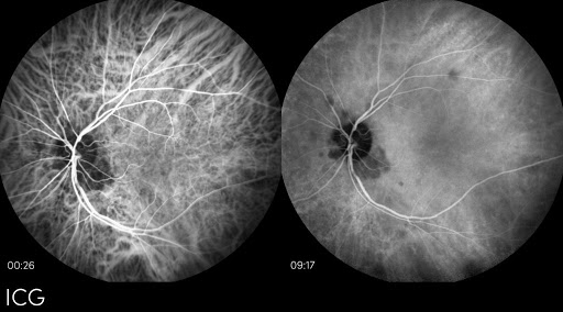
ICG Angiography Indications
Last updated: September 2026
Indocyanine green angiography (ICGA) uses a dye that binds strongly to plasma proteins and emits in the near-infrared spectrum — this lets it penetrate the retinal pigment epithelium and image the choroidal circulation in a way FFA and OCTA cannot match, which is exactly where ICGA retains an essential, largely unchallenged role.
Key indications
- Polypoidal choroidal vasculopathy (PCV): ICGA remains the diagnostic gold standard, identifying the characteristic polypoidal lesions and branching vascular network essential for distinguishing PCV from typical neovascular AMD — a distinction that materially changes treatment choice
- Occult/indeterminate choroidal neovascularization: where FFA leakage is poorly defined, ICGA can localize a "hot spot" or feeder vessel
- Central serous chorioretinopathy: demonstrates choroidal vascular hyperpermeability and dilated choroidal vessels (pachyvessels) — central to understanding CSCR as a "pachychoroid spectrum" disease
- Choroidal tumors: characterizing choroidal melanoma and hemangioma vascular patterns; helps distinguish choroidal melanoma from nevus and other simulating lesions
- Inflammatory choroidopathies: birdshot chorioretinopathy, serpiginous choroiditis, multifocal choroiditis — ICGA reveals hypofluorescent dark spots corresponding to inflammatory choroidal lesions often invisible or under-represented on FFA
- Choroidal ischemia in conditions such as pre-eclampsia/eclampsia-related choroidopathy
ICGA vs. OCTA for choroidal disease
Unlike FFA, ICGA has not been substantially displaced by OCTA — OCTA's imaging depth and its inability to show true dye leakage/hyperpermeability limit its usefulness for exactly the diseases where ICGA is most valuable (PCV, CSCR pachychoroid assessment, inflammatory choroidopathies). In current practice, ICGA and OCTA are complementary rather than competing: OCTA for structural/flow detail, ICGA for functional leakage and hyperpermeability of the choroidal circulation.
Key reference: fundamentals of ICGA in choroidal vascular disease remain consistent across recent retina imaging literature; PCV and pachychoroid spectrum disease continue to rely on ICGA as the reference standard.
📘 Want the exam-ready deep dive? This topic, and much more is covered in Ophthalmology Explorer (A High-Yield Clinical Reference for Ophthalmology Residents, Fellows & Board Exams) — available on Kindle. See all my books.
Want to know more about treatment? Read about Retina Treatment at Sadbhaav or book a consultation with Dr. Dhaval Patel.