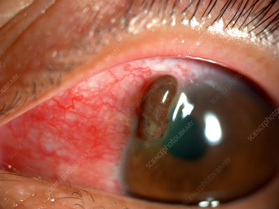
Open Globe Injury: The BETT Classification
Last updated: September 2026
Before 1996, the same word could mean completely different things in different hands — "penetrating" injury might mean a closed-globe wound into the cornea, or an open-globe wound through the entire eye wall. The Birmingham Eye Trauma Terminology (BETT) system fixed this by giving every term exactly one meaning, and it remains the international standard today.
The core principle
BETT always uses the entire eye wall (cornea + sclera) as the tissue of reference — not whichever specific tissue happens to be injured. This single rule eliminates the historical ambiguity where "corneal laceration" could mean either a full-thickness open-globe wound or a partial-thickness closed-globe injury depending on who was speaking.
The BETT injury tree
| Category | Definition |
|---|---|
| Closed globe injury | No full-thickness wound of the eye wall |
| Contusion | No wound at all; injury from the impact itself (e.g., hyphema, commotio retinae) |
| Lamellar laceration | Partial-thickness wound of the eye wall |
| Open globe injury | Full-thickness wound of the eye wall |
| Rupture | Full-thickness wound caused by blunt trauma — the eye wall fails from a sudden rise in IOP, typically at its thinnest, weakest point, which may not be at the site of impact |
| Laceration | Full-thickness wound caused by a sharp object |
| Penetrating | Single full-thickness wound, into the globe (entry only, no exit) |
| Perforating | Two full-thickness wounds — both an entry and an exit — from a single object |
| Intraocular foreign body (IOFB) | A retained foreign object causing the entry wound — see our Ocular Siderosis page for the long-term consequences of a retained metallic IOFB |
An important terminology note: per BETT, the word "blunt" describes the mechanism, while "rupture" and "contusion" describe the resulting condition — a blunt injury with a full-thickness wound is a "rupture," never a "blunt open-globe injury." This precision matters because studies that misuse this terminology (e.g., reporting a "rupture" caused by a sharp object) become impossible to interpret correctly.
The four classification variables (beyond the injury type itself)
- Type — the mechanism-based category above
- Grade — visual acuity at initial presentation
- Pupil — presence or absence of a relative afferent pupillary defect, a strong independent prognostic factor
- Zone — anteroposterior extent of the injury (Zone I: cornea/limbus; Zone II: limbus to 5mm posterior into sclera; Zone III: posterior to 5mm from the limbus) — more posterior zones carry a worse visual prognosis
The Ocular Trauma Score (OTS)
Developed from analysis of over 2,500 eye injuries in the US and Hungarian Eye Injury Registries, the OTS combines initial visual acuity with the presence of rupture, endophthalmitis, perforating injury, retinal detachment, and RAPD to predict visual outcome at 6 months. Its main clinical value is practical: giving patients and families a realistic, evidence-based expectation of visual prognosis early in the course of treatment, rather than an anecdotal guess.
Key references: Birmingham Eye Trauma Terminology (BETT), EyeWiki. | Pieramici DJ, et al. A system for classifying mechanical injuries of the eye. Am J Ophthalmol. 1997. | Kuhn F, et al. A standardized classification of ocular trauma. Ophthalmology. 1996.
📘 Want the exam-ready deep dive? This topic, and much more is covered in Ophthalmology Explorer (A High-Yield Clinical Reference for Ophthalmology Residents, Fellows & Board Exams) — available on Kindle. See all my books.
Want to know more about treatment? Read about Corneal Topography at Sadbhaav or book a consultation with Dr. Dhaval Patel.