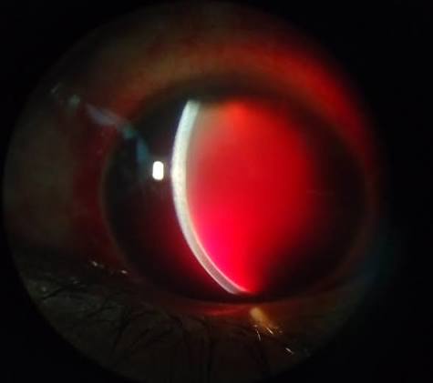
Traumatic Hyphema: Current Management
Last updated: September 2026
Traumatic hyphema — blood in the anterior chamber after blunt or penetrating injury — is common, but a genuinely important shift in the evidence base has moved practice away from a treatment that was routine teaching for decades: antifibrinolytic agents.
Why rebleeding is the central concern
Secondary hemorrhage (rebleeding), typically occurring 2-5 days after the initial injury, carries a worse prognosis than the initial bleed — the highest risk window for rebleeding is the first few days, which is why close early follow-up matters more than almost anything else in management.
Risk factors for rebleeding include:
- Higher initial hyphema grade (Grade III-IV carries substantially higher rebleed risk)
- Initial visual acuity of 6/60 or worse
- Delayed presentation/examination after injury
- Possibly higher risk in patients of African descent, per some historical series
A genuinely current shift: antifibrinolytics are no longer routine
For decades, aminocaproic acid and tranexamic acid were widely taught as standard adjuncts to reduce rebleeding risk. The evidence base has since shifted meaningfully:
- A 2022 Cochrane systematic review (23 randomized and 7 quasi-randomized trials, ~2,969 participants) found no good evidence that antifibrinolytics improve meaningful visual or complication outcomes — any effect on secondary hemorrhage rate was inconsistent, and aminocaproic acid was associated with increased nausea, vomiting, and other adverse effects
- A 2020-2021 international survey of hyphema management practices across dozens of institutions worldwide found zero institutions reported routine use of antifibrinolytics — reflecting how far actual practice has already drifted from older textbook teaching
- Topical corticosteroids and cycloplegics remain widely used and are still considered standard practice
Practical takeaway: if you were taught that antifibrinolytics are a standard part of hyphema management, know that this is now outdated — current evidence doesn't support their routine use, and most centers worldwide have already stopped using them routinely.
Standard supportive management
- Rigid eye shield (not a patch alone) at all times to prevent further trauma
- Head elevation to encourage blood to settle inferiorly, away from the visual axis and trabecular meshwork
- Activity restriction (quiet ambulation) — avoid strenuous activity
- Avoid NSAIDs, which can theoretically increase bleeding risk
- Topical cycloplegics and corticosteroids are routine at most centers
- Discourage eye rubbing
Special consideration: sickle cell disease/trait
This genuinely changes management, since sickled red blood cells obstruct the trabecular meshwork more readily than normal cells, causing IOP spikes even with a relatively small hyphema:
- Consider sickle cell screening, particularly in at-risk populations, though this is inconsistently performed in practice (only ~17% of surveyed institutions routinely test)
- Avoid carbonic anhydrase inhibitors (they can promote further red cell sickling in the aqueous) — use alternative IOP-lowering agents instead
- Have a lower threshold for surgical intervention (anterior chamber washout), since even modest IOP elevation is poorly tolerated by the optic nerve in sickle cell patients
Indications for surgical intervention
- Corneal blood staining (or high risk of it developing)
- Dangerously elevated IOP despite maximum tolerated medical therapy
- Total (8-ball) hyphema not resolving
- Sickle cell disease with sustained IOP elevation, given lower tolerance
Don't anchor on the hyphema alone
Blunt trauma severe enough to cause a hyphema is often severe enough to cause other injuries — actively examine for retinal tears, retinal detachment, vitreous hemorrhage, commotio retinae, and angle recession (which can cause delayed glaucoma years later) once the view improves enough to allow it.
Key references: Traumatic hyphema: Management, UpToDate (2024). | Gharaibeh A, et al. Medical interventions for traumatic hyphema, Cochrane Database Syst Rev. 2022. | YO Need to Know: 5 Pearls for Managing Hyphema, AAO (2024).
📘 Want the exam-ready deep dive? This topic, and much more is covered in Ophthalmology Explorer (A High-Yield Clinical Reference for Ophthalmology Residents, Fellows & Board Exams) — available on Kindle. See all my books.
Want to know more about treatment? Read about Corneal Topography at Sadbhaav or book a consultation with Dr. Dhaval Patel.