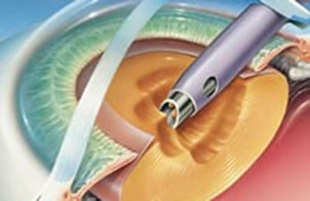
Phaco Tips & Tricks
Last updated: September 2026
These are practical, hands-on notes compiled while teaching phacoemulsification to residents and fellows — the kind of small technique adjustments that make a real difference in the OR but rarely make it into textbooks.
Download the full Phaco Tips & Tricks Manual (PDF)
Fluidics
- Master your machine's fluidics settings (bottle height/infusion pressure, aspiration flow rate, vacuum) before focusing on chopping technique — poor fluidics undermines even good instrument technique
- Higher vacuum with lower flow gives better followability for chopping; lower vacuum with higher flow suits sculpting and softer nuclei
- Watch the anterior chamber depth constantly — a shallowing chamber during phaco is an early warning sign, not just a fluidics inconvenience
Capsulorhexis
- Aim for a rhexis diameter of 5.0-5.5mm, slightly smaller than the optic diameter of most IOLs, to ensure 360° overlap of the optic edge — this reduces posterior capsule opacification and improves IOL centration
- In eyes with a mature/dense cataract and poor red reflex, trypan blue capsular staining substantially improves rhexis visualization and safety
Nucleus management
- Complete hydrodissection with visible fluid wave and free nuclear rotation before starting phaco — an incompletely mobilized nucleus increases zonular stress
- Avoid hydrodissection altogether in known or suspected posterior polar cataracts — use gentle viscodelineation instead, since the posterior capsule may already be thin or dehiscent
- Match your chop technique to nuclear density: horizontal chop for soft-to-moderate nuclei, vertical chop for dense/brunescent ones
Reading warning signs early
- A sudden deepening of the chamber, momentary pupil snap, or a fragment that unexpectedly disappears posteriorly are early signs of posterior capsule compromise — pause and reassess immediately rather than continuing
- Never chase a dropped nuclear fragment anteriorly if it has moved posterior to the capsular plane — this risks vitreoretinal traction; involve a retina specialist for posterior fragment removal if needed
For a detailed algorithm on managing an actual posterior capsule tear if one does occur, see our Posterior Capsule Tear Management page.
📘 Want the exam-ready deep dive? This topic is covered in iNotes 2027: Lens (Ophthalmology Postgraduate Exam Notes) — available on Kindle. See all my books.
Want to know more about treatment? Read about Cataract Surgery at Sadbhaav or book a consultation with Dr. Dhaval Patel.