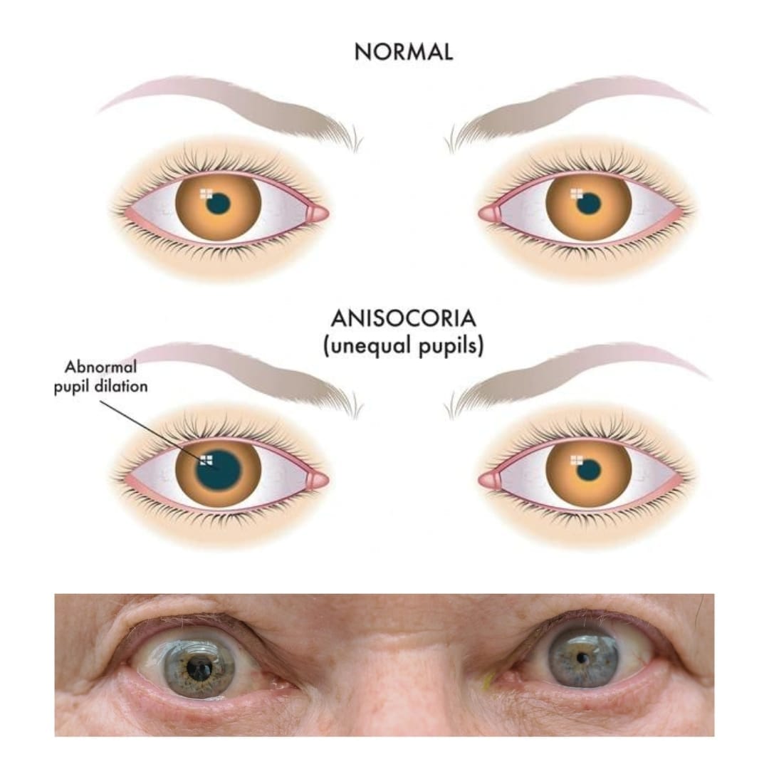
Anisocoria: A Practical Workup
Last updated: September 2026
Anisocoria (unequal pupil size) is a common referral, and most cases are benign — but the workup exists specifically to catch the rare, dangerous causes. The single most useful first step is deceptively simple: is the anisocoria greater in bright light or in darkness?
Step 1: Which pupil is abnormal?
| Anisocoria worse in | Abnormal pupil | Points to |
|---|---|---|
| Bright light | The larger pupil (fails to constrict) | Parasympathetic problem — CN III palsy, Adie's tonic pupil, pharmacologic mydriasis, iris damage/trauma |
| Darkness | The smaller pupil (fails to dilate) | Sympathetic problem — Horner syndrome, or simple physiologic anisocoria |
Physiologic anisocoria — the most common cause
Seen in up to 20% of the population: a small (usually ≤1mm), stable difference in pupil size, roughly equal in light and dark, with normal reactivity in both eyes and no ptosis. This is a diagnosis of exclusion by pattern — confirmed by old photographs showing the same asymmetry over time.
Horner syndrome: current pharmacologic testing
Apraclonidine has now replaced cocaine as the first-line confirmatory test — it's more available, doesn't require compounding, and works via denervation supersensitivity: in a normal eye apraclonidine (a weak alpha-1/strong alpha-2 agonist) has little effect, but in a sympathetically denervated eye, upregulated alpha-1 receptors cause the miotic Horner pupil to dilate and the ptosis to lift — a reversal of anisocoria confirms the diagnosis.
- A useful practical note: a negative apraclonidine test does not reliably exclude Horner syndrome — false negatives are well documented, so a strongly suggestive clinical picture with a negative test still warrants further workup
- Hydroxyamphetamine can further localize a confirmed Horner to pre- vs. post-ganglionic — but requires roughly a week's separation from the diagnostic test, so it's rarely done acutely
- In acute presentations with associated neurological symptoms, imaging should not wait for pharmacologic testing — new painful Horner syndrome demands urgent imaging (CTA/MRA of the neck) to rule out carotid artery dissection, a genuine emergency
The dilated pupil: don't miss a CN III palsy
- A dilated, poorly-reactive pupil with ptosis and/or extraocular motility restriction is a CN III palsy until proven otherwise — pupil involvement in a CN III palsy suggests compressive etiology (most importantly, a posterior communicating artery aneurysm) and requires urgent neuroimaging (CTA/MRA), not observation
- A pupil-sparing CN III palsy in a patient with vascular risk factors is more often microvascular/ischemic — but this is a diagnosis made cautiously and only after excluding compression, especially if any pupillary involvement develops on follow-up
- Adie's tonic pupil: a dilated pupil with light-near dissociation (poor reaction to light, better/tonic reaction to near) and sluggish redilation after constriction; confirmed with dilute pilocarpine (0.1%), which constricts the tonic pupil due to denervation supersensitivity but has no effect on a normal pupil
- Pharmacologic mydriasis (accidental atopine/scopolamine exposure) — the pupil fails to constrict even to strong pilocarpine (1%), which distinguishes it from a CN III palsy or Adie's pupil
Key references: Anisocoria Workup, Medscape (2025). | Adult Horner's syndrome: a combined clinical, pharmacological, and imaging algorithm. Eye. 2013. | Fierz FC, et al. Apraclonidine — an eye opener. Front Ophthalmol. 2022.
📘 Want the exam-ready deep dive? This topic, and much more is covered in Ophthalmology Explorer (A High-Yield Clinical Reference for Ophthalmology Residents, Fellows & Board Exams) — available on Kindle. See all my books.
Want to know more about treatment? Read about Corneal Topography at Sadbhaav or book a consultation with Dr. Dhaval Patel.