Optic Neuritis: Typical vs Atypical
Last updated: September 2026
Optic neuritis was once taught as a single, fairly uniform entity defined by the Optic Neuritis Treatment Trial (ONTT) — but the discovery of AQP4 and MOG antibodies has since split it into biologically distinct diseases with different prognoses and treatments. Testing for these antibodies is now part of the standard modern workup, not an optional extra.
The ONTT foundation (still relevant, but now understood as one subtype)
- Established the classic presentation: unilateral, subacute vision loss with pain on eye movement, typically in young adults
- Randomized IV methylprednisolone vs. oral prednisone vs. placebo — IV steroids accelerated visual recovery but did not change the final visual outcome; oral steroids alone (without preceding IV) were actually associated with a higher relapse rate
- Brain MRI at presentation remains the strongest predictor of conversion to multiple sclerosis — roughly 50% of ONTT participants with a first typical episode converted to MS by 15 years, with baseline MRI lesion burden driving that risk
- Key limitation to know: the ONTT was conducted before AQP4-IgG and MOG-IgG were discovered, excluded bilateral/recurrent/severe presentations, and was drawn from a population that doesn't reflect global diversity — so its findings, while foundational, don't generalize to the atypical forms below
Typical vs. atypical optic neuritis
| Feature | Typical (MS-associated) | Atypical (NMOSD / MOGAD) |
|---|---|---|
| Laterality | Unilateral | Often bilateral |
| Severity | Moderate | Often severe, poorer visual prognosis |
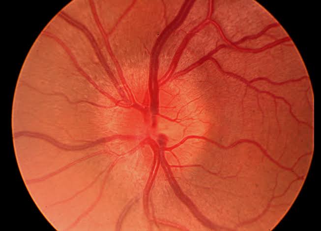
| Disc appearance | Often normal-appearing disc (retrobulbar) | Disc swelling common, especially MOGAD |
| Recurrence | Less frequent | Often recurrent |
| MRI pattern | Short-segment optic nerve lesion; brain white matter lesions typical of MS | NMOSD: longitudinally extensive, bilateral, or chiasmal involvement. MOGAD: perineural/orbital fat enhancement (optic perineuritis), often bilateral |
Current diagnostic workup
- Serologic testing for AQP4-IgG and MOG-IgG is now recommended in the standard evaluation of optic neuritis — especially in patients who don't fit the classic MS demographic (older age, or Asian/African-American/Afro-Caribbean ethnicity, where NMOSD is proportionally more common)
- Imaging has also evolved: rather than the ONTT's plain brain MRI, current practice adds dedicated orbital MRI with gadolinium and fat suppression — looking for optic nerve enhancement patterns that can themselves suggest the underlying cause (perineural enhancement favors MOGAD; chiasmal/longitudinally extensive lesions favor NMOSD)
- Spinal MRI may be warranted to look for longitudinally extensive transverse myelitis (NMOSD/MOGAD) or other demyelinating lesions
- 2024 McDonald criteria update: optic neuritis can now support an MS diagnosis at the very first demyelinating event when appropriate supportive biomarkers are present — reflecting a shift toward viewing isolated ON as often the first manifestation of MS, integrated more directly into the diagnostic framework rather than treated as a separate, "wait and see" entity
Treatment beyond the ONTT protocol
- IV methylprednisolone remains first-line acute treatment for accelerating recovery
- Plasma exchange (PLEX) is an important option for severe, steroid-refractory optic neuritis, particularly in NMOSD, where visual outcomes with PLEX after IV steroid failure can be meaningfully better than continued steroids alone
- Long-term management diverges sharply by diagnosis: MS-associated ON is managed with disease-modifying MS therapies; NMOSD and MOGAD have their own specific immunosuppressive/biologic regimens, distinct from MS treatment — getting the underlying diagnosis right isn't just academic, it changes the entire treatment pathway
Key references: Optic Neuritis in the New Millennium. TNOA J Ophthalmic Sci Res. 2024. | From the ONTT to Antibody-Mediated Optic Neuritis: Four Decades of Progress. 2026. | MOG antibody-associated optic neuritis. Eye. 2024.
📘 Want the exam-ready deep dive? This topic, and much more is covered in Ophthalmology Explorer (A High-Yield Clinical Reference for Ophthalmology Residents, Fellows & Board Exams) — available on Kindle. See all my books.
Want to know more about treatment? Read about Corneal Topography at Sadbhaav or book a consultation with Dr. Dhaval Patel.