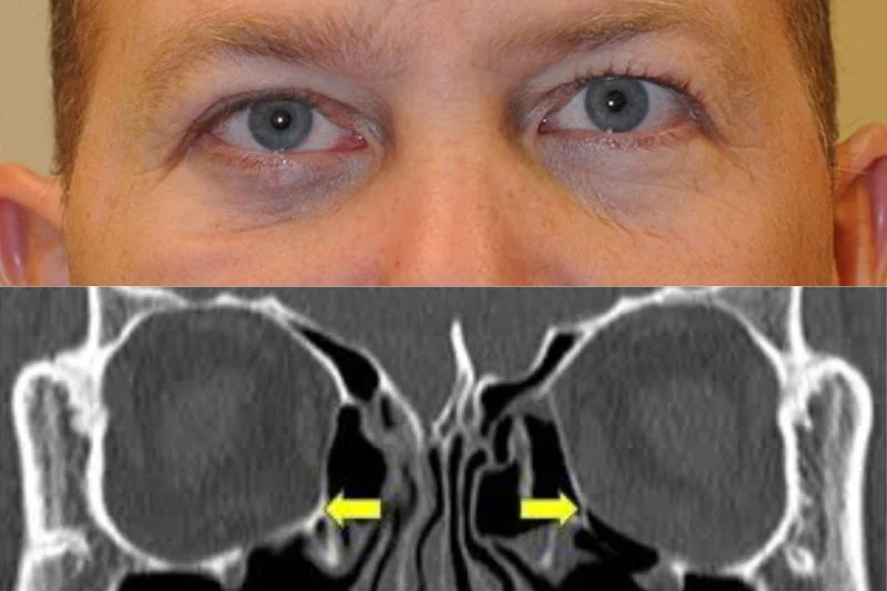
Orbital Fracture: Emergency vs Observation
Last updated: September 2026
Most orbital blowout fractures can be safely observed — but a small subset are true surgical emergencies, and confusing the two categories is the single most consequential mistake in orbital trauma management.
Mechanism
Blunt trauma to the globe or orbital rim transmits force that fractures the orbit's thinnest walls — most commonly the floor (over the maxillary sinus) or the medial wall (the lamina papyracea) — while the orbital rim itself typically remains intact. Orbital contents (fat, muscle) can herniate through the resulting defect.
The critical distinction: routine fracture vs. surgical emergency
The "white-eyed blowout fracture" is the presentation every resident must recognize immediately, because it looks deceptively mild:
- Seen classically in young patients (children and adolescents, whose more pliable bone allows a "trapdoor" fracture that snaps back into place, trapping tissue)
- Minimal external signs — little bruising or swelling ("white eye"), which is exactly what makes it easy to under-triage
- Marked restriction of vertical eye movement, out of proportion to the mild external appearance
- May be accompanied by the oculocardiac reflex: nausea, vomiting, bradycardia — triggered by the entrapped muscle, and itself an indication for urgent surgery
This entrapment scenario requires immediate surgical repair (within 24-48 hours) — delay risks ischemic muscle necrosis and permanent motility deficit, since the muscle is genuinely trapped and mechanically compromised, not just displaced.
Everything else: the 2-week observation window
For fractures without frank entrapment and oculocardiac reflex, a widely adopted evidence-based protocol (Burnstine) still guides most centers:
| Timing | Indication |
|---|---|
| Immediate | Diplopia with CT-confirmed muscle/tissue entrapment and a non-resolving oculocardiac reflex; the white-eyed blowout fracture in a young patient |
| Within 2 weeks | Symptomatic diplopia with positive forced ductions, CT-confirmed entrapment, and minimal improvement over the observation period |
| Observe (re-examine at 7-10 days) | Equivocal findings without clear entrapment |
If, after 10 days, equivocal forced ductions persist, diplopia remains in primary or downgaze, or enophthalmos exceeds 2mm with a confirmed fracture on CT, surgery should be strongly considered at that point.
Clinical indications for surgery (beyond the emergency scenario)
- Persistent diplopia in primary or reading gaze
- Positive forced duction testing (distinguishes true mechanical entrapment from muscle contusion/paresis, which will not show restriction on forced ductions)
- Enophthalmos >2mm, or large floor defects (>50% of floor area) with expected progressive enophthalmos
Radiographic findings alone are not reliable surgical indications — a 2024 systematic review found that criteria like fracture size and inferior rectus muscle rounding/displacement on CT vary significantly in their predictive reliability across studies. Clinical findings (diplopia, forced ductions, measured enophthalmos) remain the primary basis for the decision to operate, with CT used to confirm and characterize rather than to independently drive the decision.
Imaging technique
Fine-cut (1-2mm) CT in both axial and coronal planes (with sagittal reconstruction where available) is standard — coronal views best assess the floor and the posterior ledge available for implant support during repair; sagittal views help evaluate soft tissue herniation and muscle entrapment.
A practical warning for patients
Instruct patients with a suspected or confirmed orbital fracture not to blow their nose — forced air from the sinonasal tract can be pushed into the orbit through the fracture defect, causing orbital emphysema and, in severe cases, orbital compartment syndrome with risk of vision loss from acutely elevated orbital pressure.
Key references: Orbital "Blowout" Fractures: Time for a New Paradigm. Ophthalmology. 2018. | Radiographic Indications for Orbital Blowout Fracture Reconstruction: A Critical Review. 2024. | Management of orbital fractures: challenges and solutions. Clin Ophthalmol.
📘 Want the exam-ready deep dive? This topic, and much more is covered in Ophthalmology Explorer (A High-Yield Clinical Reference for Ophthalmology Residents, Fellows & Board Exams) — available on Kindle. See all my books.
Want to know more about treatment? Read about Corneal Topography at Sadbhaav or book a consultation with Dr. Dhaval Patel.