Uveitis Masquerade Syndromes
Last updated: September 2026
A "uveitis" that doesn't respond the way it should — or that keeps recurring in an older patient without a clear pattern — deserves genuine suspicion for a masquerade syndrome. Missing this diagnosis isn't just an inflammatory-control failure; in the case of intraocular lymphoma, it can be fatal.
What counts as a masquerade syndrome
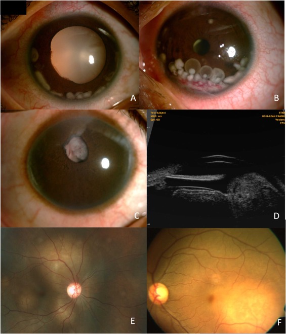
Conditions that mimic uveitis clinically but arise from an entirely different underlying process — most importantly neoplastic causes (primary vitreoretinal lymphoma, uveal melanoma, leukemia, metastatic tumors, retinoblastoma in children), but also non-neoplastic mimics. These account for roughly 2.5% of presumed uveitis cases in tertiary referral settings — uncommon, but not rare enough to ignore.
Primary vitreoretinal lymphoma (PVRL): the one you cannot afford to miss
- Peak incidence in older adults, 50-70 years — a genuinely useful epidemiologic clue, since new-onset "uveitis" in this age group deserves higher suspicion than in a young adult
- Classic presentation: vitritis, anterior chamber cells, and creamy yellow-white subretinal/sub-RPE infiltrates — often initially treated as chronic posterior or panuveitis
- Less common presentations: hyphema, pseudohypopyon, exudative retinal detachment, pseudovasculitis — a genuinely broad masquerading repertoire
- PVRL is a subtype of primary CNS lymphoma — a meaningful proportion of patients have or will develop CNS involvement, so brain MRI and neuro-oncology input matter once suspected
Why diagnosis is so often delayed
Diagnosis is frequently delayed up to a year after symptom onset, for a specific, avoidable reason: corticosteroids are lympholytic. Treating presumed uveitis with steroids can transiently improve the clinical picture (reinforcing the wrong diagnosis) while simultaneously causing fragile lymphoma cells to lyse — which can render a subsequent vitreous biopsy falsely negative and further delay the correct diagnosis. Practical rule: maintain a low threshold for diagnostic vitreous biopsy in atypical, older-onset, or poorly-responsive uveitis before committing to prolonged steroid treatment.
Diagnostic tools (current standard)
- Vitreous biopsy via pars plana vitrectomy remains the primary diagnostic method — ideally obtaining at least 1mL of undiluted vitreous, with the pathologist informed and prepared in advance since cytological yield is often low and cells are fragile
- Cytology alone has limited sensitivity due to the small quantity and fragility of cells in vitreous samples — repeat biopsy is not uncommon
- MYD88 L265P mutation testing — a gain-of-function mutation causing constitutive NF-κB activation, present in roughly 69-89% of PVRL cases; PCR-based testing on vitreous/aqueous samples is an increasingly important molecular diagnostic adjunct to cytology alone
- IL-10/IL-6 ratio in vitreous or aqueous — an IL-10/IL-6 ratio greater than 1, or markedly elevated IL-10 alone, supports lymphoma over inflammatory uveitis, though false-positive (~11%) and false-negative (~23-30%) rates mean this is supportive evidence, not a standalone diagnostic test
- Immunoglobulin heavy chain (IgH) and T-cell receptor (TCR) gene rearrangement studies provide molecular confirmation of B-cell or T-cell clonality respectively
- Multiparametric flow cytometry of aqueous or vitreous fluid, when available, can identify a monoclonal B-cell population directly
Practical red flags that should prompt considering a masquerade
- "Uveitis" in a patient over 50 without a clear prior inflammatory pattern
- Poor or incomplete response to appropriately dosed corticosteroids
- Absence of pain and posterior synechiae (more typical of neoplastic disease than true inflammatory uveitis, though not absolute)
- Known systemic malignancy or lymphoma history
- Unilateral presentation with an atypical inflammatory pattern
Key references: Neoplastic Masqueraders of Uveitis. Retina Today. 2025. | Uveitis Masquerade Syndromes, EyeWiki. | Rothova A, et al. Causes and clinical manifestations of masquerade syndromes in intraocular inflammatory diseases. Retina. 2021.
📘 Want the exam-ready deep dive? This topic, and much more is covered in Ophthalmology Explorer (A High-Yield Clinical Reference for Ophthalmology Residents, Fellows & Board Exams) — available on Kindle. See all my books.
Want to know more about treatment? Read about Corneal Topography at Sadbhaav or book a consultation with Dr. Dhaval Patel.