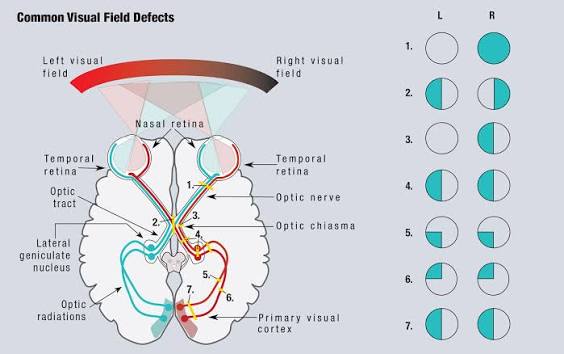
Visual Field Defect Localization
Last updated: September 2026
The shape of a visual field defect is a map of where along the visual pathway the lesion sits — this is genuinely one of the highest-yield localization skills in neuro-ophthalmology, both for exams and for real triage decisions.
The core localization rules
| Pattern | Localization |
|---|---|
| Purely unilateral field defect (one eye only) | Pre-chiasmal — retina or optic nerve of that eye |
| Bitemporal (heteronymous) hemianopia | Chiasmal — classically pituitary tumor or craniopharyngioma, compressing the crossing nasal retinal fibers |
| Homonymous hemianopia (same side, both eyes, respecting vertical midline) | Post-chiasmal — anywhere from the optic tract to the occipital cortex |
Localizing further within homonymous defects: the congruity rule
The more congruent (identical in both eyes) an incomplete homonymous hemianopia is, the more posterior the lesion.
- Optic tract lesions — typically produce an incongruous homonymous defect (about 50% of tract lesions show incongruity), often with a relative afferent pupillary defect in the contralateral eye since the tract carries more fibers from one eye than the other at this level
- Optic radiation lesions — the large majority of incongruous defects arise here (a more anterior location than the tract logic might suggest, but the anatomy is complex); a temporal lobe lesion (Meyer's loop) produces a superior quadrantanopia ("pie in the sky"); a parietal lobe lesion produces an inferior-denser homonymous defect
- Occipital lobe lesions — produce the most congruous defects (about 84% congruous in one series); a complete homonymous hemianopia, however, has essentially no further localizing value beyond confirming a post-chiasmal, non-tract location — completeness alone can't distinguish a radiation lesion from an occipital one
Important caveat: the congruity rule is a strong tendency, not an absolute — only incomplete hemianopias can be meaningfully described as congruous or incongruous at all.
Special patterns worth recognizing
- Junctional scotoma — an ipsilateral central field defect combined with a contralateral superotemporal defect; occurs from a compressive lesion at the junction of the optic nerve and chiasm, where the crossing inferonasal fibers of the contralateral eye loop forward into the ipsilateral nerve (Wilbrand's knee) before crossing — a classic, specific localizing sign
- Macular sparing — occurs with occipital lobe lesions due to dual blood supply to the occipital pole (middle cerebral artery collateral in addition to posterior cerebral artery) — a useful clue pointing specifically to the occipital cortex rather than more anterior post-chiasmal structures
- Riddoch phenomenon — some patients with apparently complete homonymous hemianopia retain conscious perception of moving stimuli in the "blind" hemifield, thought to reflect a preserved direct pathway from the lateral geniculate nucleus to visual area V5 that bypasses the primary visual cortex — a genuinely interesting example of residual visual function surviving apparent complete field loss
Practical framework
- Does the defect cross the vertical midline? If yes → pre-chiasmal (unilateral) or chiasmal (bitemporal); if no → post-chiasmal (homonymous)
- For homonymous defects, assess congruity to estimate anterior-to-posterior location
- Look specifically for junctional scotoma or macular sparing — both carry outsized localizing value relative to how easy they are to check for
- Correlate with imaging — MRI brain/orbits is the definitive next step once the field pattern narrows the differential
Key references: Homonymous Hemianopsia, StatPearls (2024). | Neuro-Ophthalmology for Neuroradiologists. AJNR. | Neuro-ophthalmology Illustrated, Chapter 3: Visual Fields, Stanford.
📘 Want the exam-ready deep dive? This topic, and much more is covered in Ophthalmology Explorer (A High-Yield Clinical Reference for Ophthalmology Residents, Fellows & Board Exams) — available on Kindle. See all my books.
Want to know more about treatment? Read about Corneal Topography at Sadbhaav or book a consultation with Dr. Dhaval Patel.