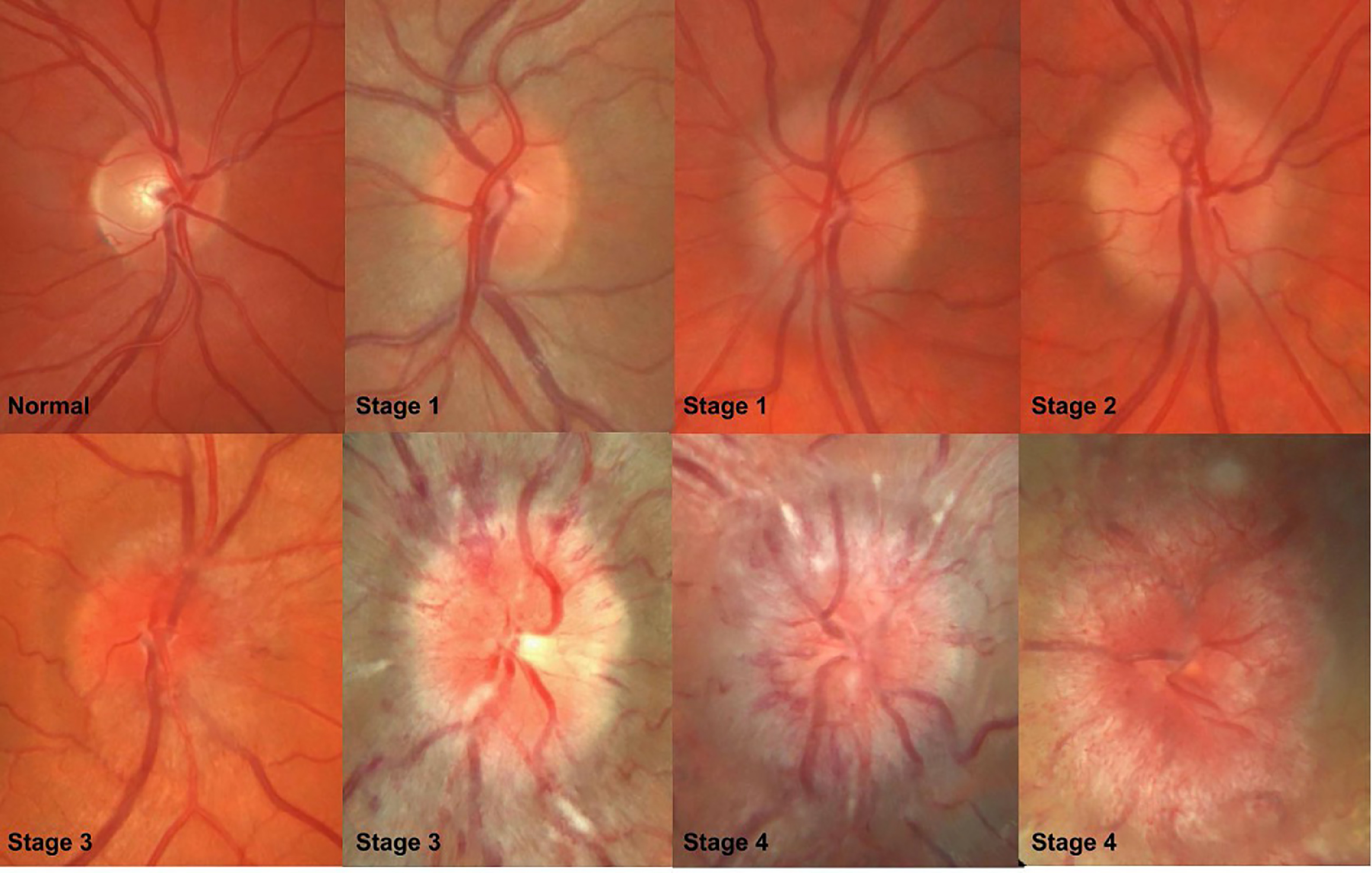
Papilledema vs Pseudopapilledema
Last updated: September 2026
Distinguishing true papilledema from pseudopapilledema matters enormously — misdiagnosing pseudopapilledema as papilledema leads to unnecessary lumbar punctures and neuroimaging; missing true papilledema risks missing a mass lesion, venous sinus thrombosis, or idiopathic intracranial hypertension. OCT has become central to making this distinction reliably.
Definitions
- Papilledema — optic disc swelling specifically due to raised intracranial pressure; the retinal nerve fiber layer itself is edematous
- Pseudopapilledema — anomalous elevation of the optic disc without true nerve fiber layer edema; most commonly caused by optic disc drusen, but also small crowded discs, tilted discs, and other congenital anomalies
Clinical clues favoring true papilledema
- Bilateral (true papilledema from raised ICP is almost always bilateral; pseudopapilledema can be unilateral or bilateral)
- Peripapillary hemorrhages, cotton wool spots, and choroidal folds are strong supporting signs when present
- Obscured disc margins with a blurred, indistinct vessel pattern crossing the disc edge
- Associated symptoms: headache, transient visual obscurations, pulsatile tinnitus, diplopia (especially CN VI palsy from raised ICP)
OCT: the current primary distinguishing tool
| OCT finding | Papilledema | Pseudopapilledema (ODD) |
|---|---|---|
| Internal disc contour | Smooth | "Lumpy-bumpy" irregular contour |
| Peripapillary RNFL thickness | Thickened, typically all quadrants (nasal quadrant has highest diagnostic value) | Normal or thin |
| Macular ganglion cell-inner plexiform layer | Normal early; thins later if chronic | Can be thinned if drusen compress axons directly |
| Hyperreflective subretinal material | Absent | May show hyperreflective bands/lumps corresponding to buried drusen |
SD-OCT has shown higher sensitivity than B-scan ultrasonography in several recent comparative series, though the two remain complementary in practice — ultrasound is excellent at directly visualizing calcified drusen (hyperechoic with shadowing) and can also assess optic nerve sheath diameter (a diameter >3.3mm supports raised ICP), while OCT gives detailed structural quantification of the nerve fiber layer itself.
Other supporting tools
- Fundus autofluorescence — buried optic disc drusen are autofluorescent and light up well before they become visible clinically, making this a fast, non-invasive screening step
- Fluorescein angiography — true papilledema shows early diffuse disc hyperfluorescence with late leakage (from blood-retina barrier breakdown); this leakage pattern is absent in pseudopapilledema
- OCT angiography (OCTA) — an active area of research; papilledema tends to show higher peripapillary flux index and perfusion, particularly nasally, though diagnostic accuracy is currently only moderate as an adjunct rather than a standalone test
A practical caveat
Optic disc drusen do not exclude a patient from also developing true papilledema later — a patient with known ODD who develops new peripapillary hemorrhages or subjective symptoms still deserves reassessment, not automatic reassurance based on their prior drusen diagnosis. Conditions like Chiari malformation can coexist with ODD, and distinguishing genuine ICP elevation from baseline drusen-related disc elevation in that setting specifically warrants multimodal imaging (OCT + autofluorescence + ultrasound) rather than clinical impression alone.
Key references: Pseudopapilledema, EyeWiki (2025). | Validity of OCTA in the differential diagnosis of papilledema and pseudopapilledema. 2025. | A field guide to optic disc drusen. Modern Retina. 2026.
📘 Want the exam-ready deep dive? This topic, and much more is covered in Ophthalmology Explorer (A High-Yield Clinical Reference for Ophthalmology Residents, Fellows & Board Exams) — available on Kindle. See all my books.
Want to know more about treatment? Read about Corneal Topography at Sadbhaav or book a consultation with Dr. Dhaval Patel.