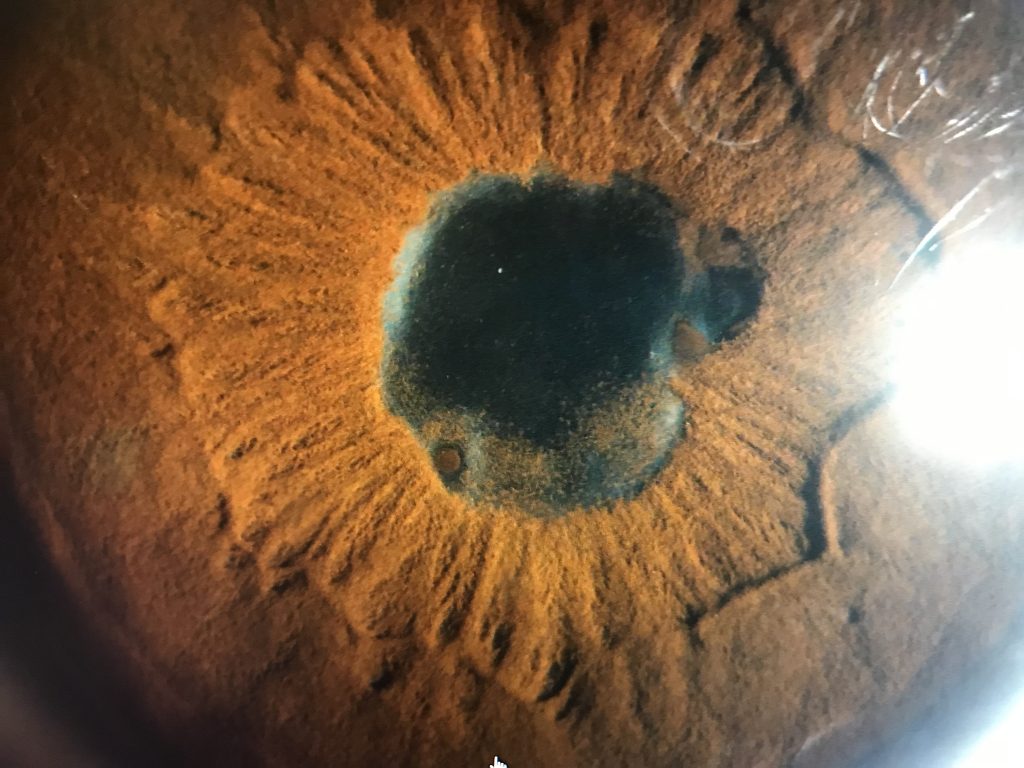
Uveitis
Last updated: September 2026
Causes of unilateral uveitis
- Sarcoidosis
- Postsurgical uveitis
- Intraocular foreign body
- Parasitic disease
- Acute retinal necrosis
- Behçet's disease
Causes of panuveitis
- Syphilis
- Sarcoidosis
- Vogt–Koyanagi–Harada syndrome
- Infectious endophthalmitis
- Behçet's disease
Causes of granulomatous inflammation in the eye
- Sarcoidosis
- Sympathetic ophthalmia
- Uveitis associated with multiple sclerosis
- Lens-induced uveitis
- Intraocular foreign body
- Vogt–Koyanagi–Harada syndrome
- Syphilis
- Tuberculosis
- Other infectious agents
Major fluorescein angiographic findings in uveitis
- Cystoid macular edema
- Subretinal neovascular membranes
- Disc leakage
- Late staining of retinal vessels
- Neovascularization of retinal vessels
- Retinal vascular capillary dropout and reorganization
- Retinal pigment epithelium perturbations
granulomatous uveitis is characterized by nodular collections of epithelioid cells and giant cells surrounded by lymphocytes, whereas nongranulomatous uveitis is characterized by diffuse infiltration of lymphocytes and plasma cells.
The current standard: SUN (Standardization of Uveitis Nomenclature) classification
Rather than relying on ad hoc descriptions, uveitis is now classified using the SUN Working Group's anatomic framework — this matters because it's now the universal language used in the literature, in referrals, and in tracking a patient's course over time.
Anatomic classification (based on site of primary inflammation, not just complications)
- Anterior uveitis — primary site: anterior chamber
- Intermediate uveitis — primary site: vitreous (includes pars planitis)
- Posterior uveitis — primary site: retina or choroid
- Panuveitis — significant inflammation in the anterior chamber, vitreous, AND retina/choroid simultaneously
A key SUN principle worth remembering: classification is based on the anatomic location of inflammation, not on structural complications (like cystoid macular edema or cataract) that might result from it — a common source of confusion.
Standardized grading scales
| Grade | Anterior chamber cells (cells per field) | Vitreous haze |
|---|---|---|
| 0 | <1 | None |
| 0.5+ | 1-5 | Trace — slight blurring of optic disc margin |
| 1+ | 6-15 | Mild |
| 2+ | 16-25 | Moderate |
| 3+ | 26-50 | Marked |
| 4+ | >50 | Severe — obscures optic disc |
This ordinal 0 to 4+ grading is used consistently for anterior chamber cell count, anterior chamber flare, and vitreous haze — giving a reproducible way to document severity and track improvement or worsening visit-to-visit, and to define standardized outcomes ("inactive" disease, "improvement," "worsening," "corticosteroid-sparing" success) for both clinical practice and research reporting.
Why this standardization mattered
Before SUN, agreement among uveitis experts on diagnosis was only moderate at best (kappa as low as 0.23-0.39 for some conditions) — essentially close to chance agreement for certain diseases. The SUN Working Group's more recent "SUN II" project has since developed formal classification criteria for 25 of the most common uveitic entities using rigorous case collection and machine learning validation, achieving 93-99% accuracy depending on anatomic class — a genuinely major step in making uveitis diagnosis and research reproducible across centers.
Key references: Jabs DA, et al. Standardization of Uveitis Nomenclature for Reporting Clinical Data. Am J Ophthalmol. 2005;140:509-516. | SUN Working Group. SUN II Classification Criteria (multiple entity-specific papers). Am J Ophthalmol. 2021;228.
📘 Want the exam-ready deep dive? This topic, and much more is covered in Ophthalmology Explorer (A High-Yield Clinical Reference for Ophthalmology Residents, Fellows & Board Exams) — available on Kindle. See all my books.
Experiencing symptoms like these? Learn about our Eye Department or book a consultation with Dr. Dhaval Patel.